In Part 1 we showed you what a glioblastoma cell looks like, and astrocytes, which perform many important brain functions. Next, we look at the neuron, the most commonly known brain cell, but there is still a lot you probably do not know about it.
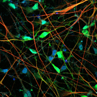
Neurons are the great communicators of the brain, responsible for transmitting signals between/within brain regions, and to glands and muscles. When you lift your arm, it is because neurons sent an electrochemical pulse along a chain of neurons from the brain to the muscles in your arm.
The brains of mammals contain 100-200 billion neurons depending on the species. Typically, neurons are divided into three types: sensory neurons, which process stimuli from our senses like touch and smell; motor neurons, that send and receive signals to control muscle and gland activity, and interneurons that connect neurons together.
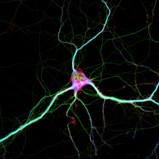
Neurons are the fundamental cells in the brain and are made up of multiple parts: dendrites carry information from other neurons to the soma, or cell body; the nucleus in the soma contains the neuron’s DNA, and is also where proteins are made to be transported throughout the dendrites and the axon.
Axons are long tentacle-like arms that stretch from the nuclei of neurons to other neurons. Signals travel from the cell body up the axon to synapses, where the signals cross narrow gaps via neurotransmitters. The signal is received by the dendrite of another neuron where it is passed to the cell body, and the chain continues.
Most axons are tiny but some grow very long. The axons of the sciatic nerve are the longest in the human body, running from the base of the spine to the big toe of each foot.
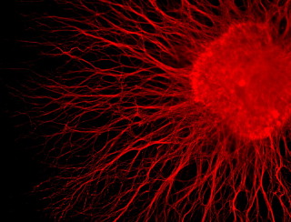
The word dendrite comes from the Greek word for tree and you can see why. Dendrites project outward from the cell body into complex patterns almost like branches. Along the branches are synapses where signals pass from the axons of other neurons.
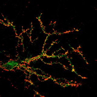
This image shows the dendritic tree of a neuron isolated from the hippocampus and grown in vitro (in a dish), with its synapses marked in red. Since this is one dendrite of one neuron, and there are billions of neurons in a single human, you can imagine how many signals are being sent across synapses in your brain right now.
Now you know the basic parts of the neuron: the dendrites, axon, nucleus and synapses. The neuron is microscopic–you cannot see it with the naked eye. But neuroscientists study structures even smaller than the neuron. They use tools like electron microscopes to take images of the building blocks of neurons, such as the organelles.
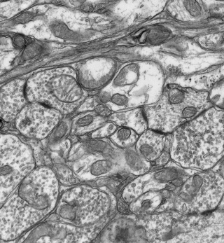
Organelles are structures that perform specific tasks within a cell, like how our organs perform certain functions to keep us alive and healthy. This image shows organelle components of synapses. They help carry neurotransmitters to the synaptic gap, which they cross to reach receptors on the other side. Understanding their mechanism will help us develop therapies for conditions where there is synaptic misfiring.
Electron microscopes make studying these tiny organelles possible. They use elections as a source of illumination, which have much smaller wavelengths than photons and so can produce images of small objects at higher resolution than a traditional light microscope. This image was taken by Alexandra Brignall at the lab of JF Cloutier of The Neuro.
So far, we have looked at brain cells types and their components. In Part 3, we look at more complex structures, and how they help us sense and understand our surroundings.