The last part of our series looks at the different structures of the brain, its mechanisms of function. In neuroscience, it is not enough to look at single cells in isolation. The brain is a system, and scientists need to understand how different cells interact to perform their functions.
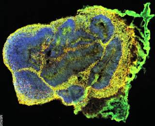
The Early Drug Discovery Unit at The Neuro is growing brain organoids to study neurological disease. These “mini-brains” measure up to four millimetres and are made of similar cell types like a real brain, including neurons and glial cells.
Brain organoids provide the closest possible model to patient’s brains, offering a precise and innovative approach to investigate disease mechanisms and accelerate discovery of new therapeutics. The EDDU uses these organoids to study neurodegenerative disorders such as Parkinson’s, ALS as well as neurodevelopmental disorders such as microcephaly.
A large part of the brain’s function is interpreting information from our senses to construct the world around us. Light entering our eyes, sounds from our ears, and the pressure of touch all need to be detected by sensory neurons and processed by different parts of the brain.
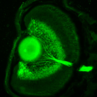
The image comes from the lab of Ed Ruthazer at The Neuro, where they study the development of the visual system, among other things. It is the eye of a Xenopus tadpole expressing green fluorescent protein in the ganglion cells of the retina, which in vertebrates form the projection from the eye to the brain. The optic nerve can be seen exiting the eye on the right.
We move from sight to smell and the olfactory bulb: a part of the brain that receives scent signals from our nose and sends them to the amygdala for further processing. In most vertebrates, the olfactory bulb is in the front of the brain but in humans it is on the inferior or bottom. The size of this structure generally corresponds to smell sensitivity.
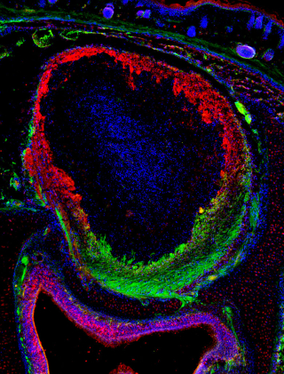
This is a tissue section of a mouse olfactory bulb showing two populations of olfactory receptor neuron axons (red and green) innervating different regions of the olfactory bulb. The photo comes from the lab of Jean-François Cloutier, where researchers study how neurons are born, project axonal processes, and form synaptic connections during neurodevelopment.

We hope you enjoyed your journey through the brain. Of course, this series covers just a few aspects of this incredibly complex organ. We encourage you to learn more about the brain and ways you can keep yours in top condition. Follow The Neuro on social media and our website for the latest developments in neuroscience research and patient care.