Next week will mark one year since the creation of the Tanenbaum Open Science Institute, crystalizing the MNI’s transformation into the world’s first fully open science institute; an endeavour made possible thanks to a generous donation from the Lawrence and Judith Tanenbaum Family Foundation. During this time, we have worked hard in a number of areas; building an open repository of patient samples, planning the necessary IT infrastructure to handle large scale data sharing, forging unique and promising open drug discovery projects with partners in industry and hiring a number of researchers with impeccable open science credentials.
It is fitting that this year culminates with the launch of our own open science publishing platform, MNI Open Research operated on our behalf by F1000. To me, it is crucial that our work, whether it’s a positive or negative result, a novel method or a description of an important new software tool or dataset, is shared quickly and transparently. This platform, which publishes findings and associated data from MNI researchers and their collaborators immediately, which will subsequently undergo open peer review, will help our mission to accelerate the discovery of cutting-edge treatments for patients suffering from neurological diseases.
Today is another step on our journey to become an open science institute, as MNI Open Research publishes its first articles. The platform launches with three articles from our researchers and their collaborators, with more on the way.

The first is a research article from my own lab in which we describe an antibody that can specifically detect the polyalanine-tract portion of proteins encoded by genes known to be pathogenic upon an expansion of their normal trinucleotide repeat numbers. A polyalanine-tract expansion is known to underlie a specific form of muscle disorder known as oculopharyingeal muscular dystrophy, and polyalanine-tracts were also predicted and observed to occur in neurodegenerative diseases associated with CAG-trinucleotide expansions (e.g. spinocerebellar ataxia and Huntington’s disease). The antibody reported in the article revealed abnormal polyalanine-tract signals in tissues and models of these
diseases. Antibodies are valuable tools and information regarding the validation of the antibody was provided so that it could be reproduced. Often the publication of new antibodies is poorly documented (e.g. nature of the antigen, validating conditions). Alternatively, some new potentially valuable antibodies may remain unreported if the global impact of manuscripts in which they were developed and used, do not warrant publication in a specialized journal. It seems unambiguous to me that the MNI Open Research operated by F1000 offered an excellent opportunity to report this antibody recognizing polyalanine-tracts. As the antibody only revealed polyalanine-tracts of pathological length, and not those from shorter non-pathological length, such a tool could prove valuable to test the contribution of polyalanine in other neurological conditions.
 The second is a detailed method developed by Brigitte Ritter (Boston University) in collaboration with colleagues from the Department of Cell Biology and the Kavli Institute for Neurosciences at Yale University when she was a member of Peter McPherson’s group at the MNI. It outlines a protocol to reduce the expression of individual or multiple proteins simultaneously in neurons, to the extent that they are not detectable by Western blot. The approach involves embedding small hairpin RNAs into microRNAs tagged to a fluorescent protein, transferring this combination into a lentivirus, which delivers this package into neurons. The protocol is quick and, unlike some other methods, shows little to no toxicity in neuronal cell cultures. The clear step-by step instructions laid out in this article should facilitate the uptake of this technique by other labs.
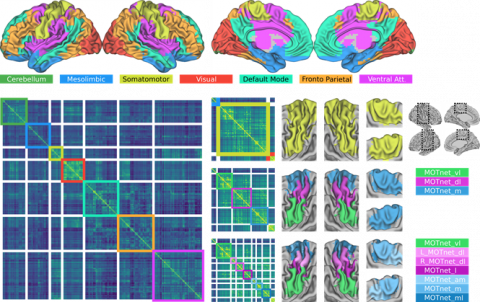 In the third, a data note, from Sebastian Urchs, a PhD student co-supervised by Alan Evans in the MNI’s Brain Imaging Centre and Pierre Bellec’s group at Université de Montréal, provides a segmentation of the brain into stable regions based on functional connectivity, across a wide range of resolutions. The template (which they call a multiresolution intrinsic segmentation template, or MIST) was created using a large open dataset from Harvard University, showing that we can benefit from the open data practices of others just as we expect others to benefit from the data we are making open. Their article makes full use of the capabilities of the web; the authors created an impressive interactive dashboard that enables readers to visually explore the hierarchical organization of these brain segmentations, and several of the figures within the online version of the article are interactive. These interactive media will enable readers to explore the MIST parcels in all their complexity, without the limitations inherent to traditional, static figures. Such advances in visualization are important for the open sharing of research outcomes in the context of big data explosion in neuroscience.
All three groups now await the comments from invited expert referees using an author-led post-publication peer review model based on that used on F1000Research. These will not be for our eyes-only; their signed reports will be published alongside our articles for everyone to read, a level of transparency that will help readers better assess our work, encourage constructive criticism, and give deserved credit to the reviewer for the time, effort and expertise they put into their report. If you are interested to hear what the reviewers think of our work, there is a tracking button on the article, which will keep you abreast of the review process, as well as notify you if or when we publish a new version of our articles. This level of transparency will help increase early career researchers’ expertise about the academic publication process. Being able to see the whole publication process enables researchers to develop and sustain the skills needed to effectively share their research to the wider community.
We decided to publish on this platform so that our data, findings and methods are quickly and easily used by others in their own efforts to understand neurological diseases. So, for those interested in neuroscience, we encourage you to sign up for article alerts on the platform (sign up is available on each article page) in order to fully benefit from the early access to results it provides, and look forward to potential new collaborations with those who wish to make use of our open scientific resources.
Source: F1000 Blog