
Age/sex: 11-year-old female
Size: 21.1 x 8.6 x 5.2 cm
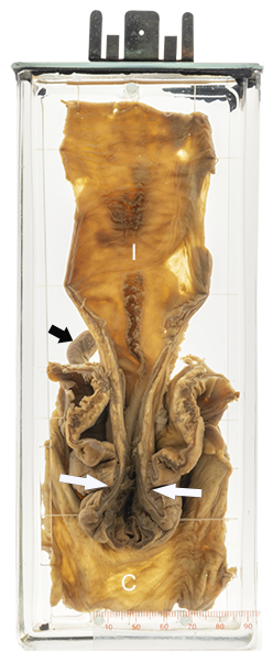
The specimen consists of the terminal ileum (I) and cecum (C). The tip of the appendix can be seen behind at the left (short arrow). The last part of the ileum has telescoped into the cecum past the ileocecal valve (long arrows). The lumen in this segment is compressed; that of the more proximal ileum is dilated.
Intussusception
Intussusception occurs when one part of the bowel folds inside an adjacent part, much like two parts of a telescope. The most common site is at the ileocecal valve, with the distal ileum extending into the cecum. The cause is not always clear. In some cases, normal intestinal contractions (peristalsis) “pulls” a polyp and the adjacent bowel wall into the distal bowel. However, such a leading lesion is not always seen.
The telescoping of the two bowel segments can lead to compression of their blood vessels and decreased blood flow (ischemia). This in turn can lead to death (necrosis) of the intussuscepted bowel mucosa followed by bleeding (classically described as “red currant jelly stool”). Symptoms include abdominal pain, nausea, and vomiting.
The condition is often successfully treated by enema, which pushes the inner bowel loop from the outer one. However, if this is not successful or if the condition progresses to mucosal necrosis (bloody stool), surgery may be required.
The disease was first described by the Dutch physician Paul Barbette in 1674. In 1876, Hirschsprung first reported the technique of enema reduction.
Below: Diagrammatic illustration of intussusception.
Source: Remesz, O. (2013). Intussusception. Wikimedia Commons.
