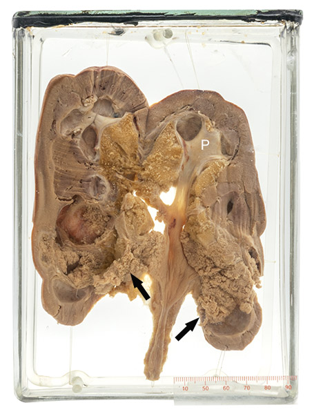
Age/sex: unknown
Size: 19.3 x 13.8 x 6.3 cm
The specimen has been cut in its mid-portion to show the two kidney halves. A small amount of normal smooth pelvis/calyx can be seen (P). The rest is covered by a finely granular tumor (arrows); compare with the solid appearance of the carcinoma in Specimen 25. The granularity reflects tumor growth as papillae.
Papillary carcinoma
Most renal tumors arise from epithelium that lines small tubules in its cortex (see Specimen 25). Papillary carcinoma originates in the lining of the urinary collecting system (the calyces and the pelvis). A papilla is defined as a finger-like growth consisting of a central fibrovascular core covered by epithelium. Because each papilla is separate from its neighbor, groups of them (such as in a tumor) result in an appearance similar to a thick knitted carpet.
Renal papillary carcinomas are often associated with papillary tumors in the bladder and ureter. Many are “low grade”, i.e., they do not invade the underlying tissue or spread elsewhere in the body (metastasize). However, larger tumors, such as the one in this specimen, often invade the adjacent renal tissue and have a worse prognosis.
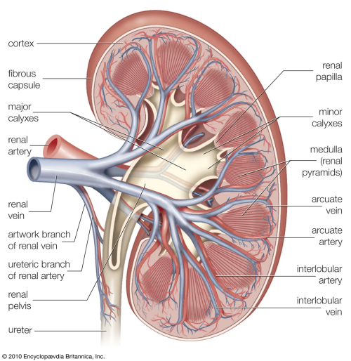
Below: A sagittal cross-section of a normal kidney.
Source: Human Kidney. Encyclopaedia Britannica. https://www.britannica.com/science/ kidney/images-videos#/media/1/317358/99762