
Age/sex: 13-year-old male
Size: 25.5 x 13.0 x 11.5 cm
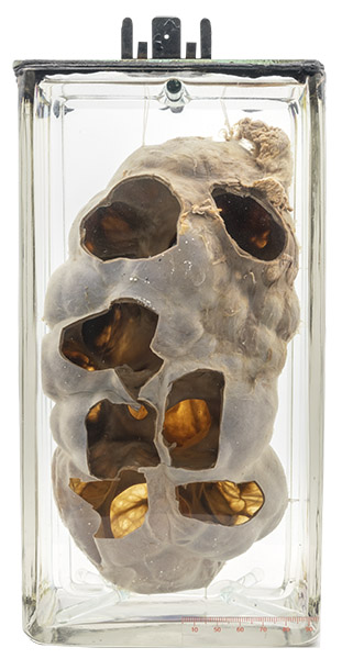
The specimen is hardly recognizable as a kidney, with the renal cortex measuring only 1-2 mm in thickness. Its interior – demonstrated well by the holes cut in the kidney surface – consists of markedly dilated calyces (the urine collecting part of the kidney).
Hydronephrosis
Hydronephrosis is defined as dilatation of the renal pelvis and calyces as a result of obstruction to urine flow. The obstruction can be anywhere in the urinary tract from the kidney to the urethra. The most common causes are ureteral reflux (in children), cancer, stones, and prostatic hyperplasia. Rarely, the etiology is an abnormality originating outside the urinary tract, such as the aberrant artery in the illustration below. This was also the cause in the specimen displayed, although the artery was not included in the preparation.
The kidneys receive their blood supply from the aorta, usually one branch on each side. However, additional branches are seen in about one-third of normal people. Sometimes, one of these branches passes in front of the ureter near the ureteropelvic junction (PUJ), compressing it and causing partial obstruction to urine flow. Over time, this leads to increased pressure in the renal pelvis, causing it to dilate and the renal cortical tissue to atrophy.
Below: Illustration showing an aberrant renal artery compressing the pelvi-ureteric junction and resulting in hydronephrosis.
Source: Panos, E. (2019). Hydronephrosis and aberrant renal artery. Critical Care Sonography. https://www.criticalcare-sonography.com/2019/11/20/congenital-hydronephrosis-and-aberrant-renal-artery/
![Hydronephrosis and aberrant renal artery [PNG]. Critical Care Sonography](https://www.mcgill.ca/medicalmuseum/files/medicalmuseum/26_reference.png)