
Donor: Dr. M.B. MacKrugir
Date: 1949
Size (H x W x D cm): 15 x 11 x 5
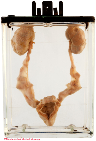
The right kidney shows innumerable small cysts. Both ureters are dilated and tortuous and the bladder wall is hypertrophied (the result of a posterior urethral valve, which is not seen in the specimen as mounted).
History: Four hour-old male infant. History unknown.
Comment: The definitive kidney develops from metanephric mesenchyme (blastema) located in the hindgut. It is connected by a ureteric bud to the excretory (cloacal) outlet. This bud forms the ureter, pelvis and collecting ducts; the blastema forms the nephron from glomerulus to the distal convoluted tubule. Multicystic dysplastic kidney is thought to occur as a result of abnormal interaction between the blastematous tissue and ureteric bud, resulting in failure of the former to develop and communicate with the collecting ducts. This is manifested grossly by renal cortical cysts and microscopically by primitive appearing ducts and glomeruli with intervening fibrous, muscle and cartilagenous tissue.
Cystic dysplasia occurs in about 1 in about 3500 births. Ninety per cent of cases are related to obstruction and 10% are hereditary. When the abnormality is isolated and confined to one kidney, symptoms and complications are usually absent. In fact, the affected kidney often involutes during childhood.