
Donor: Royal Victoria Hospital
Date: 1940
Size (H x W x D cm): 20 x 15 x 8
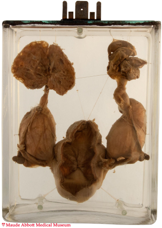
Both ureters are tortuous (seen best on the left side in a posterior view) and greatly enlarged (diameter of the lower left ureter − 3.5 cm). Both kidneys show hydronephrosis and partial replacement of the parenchyma by cysts (particularly on the right). The bladder is dilated, suggesting obstruction. However, although the urethra is not included with the specimen, its descriptive card states that no obstruction was seen, and a urethral valve or other physical obstruction was presumably absent.

History: Male infant. History unknown.
Comment: Mega-ureter is defined as abnormal dilatation of one or both ureters. It can be the result of distal obstruction (“secondary” mega-ureter), in which case it is usually caused by abnormalities of the bladder or urethra (e.g., posterior urethral valve; see specimen 34). More commonly, the condition is “primary”, either with or without associated vesico-ureteral reflux.
Refluxing primary megaureter is a result of an abnormal vesico-ureteric junction, which interferes with the normal anti-reflux mechanisms and allows urine to flow from the bladder toward the kidney. Non-refluxing primary mega-ureter does not have this complication. It is thought to be the more common cause of primary megaureter. The cause is unknown.
Patients are often asymptomatic in all three types. When present, symptoms are related to complications of urinary stasis/reflux, such as infection (pyelonephritis) or stone formation (nephrolithiasis). Other urinary tract abnormalities, such as seen in this case, are present in some patients.