
![]() Descriptive Card
Descriptive Card ![]() Log Book Entry
Log Book Entry
Rodin Number: 11
E Number: 146
Donor: Howard and Osler
Date: unknown
Size (H x W cm): 15 x 12.5
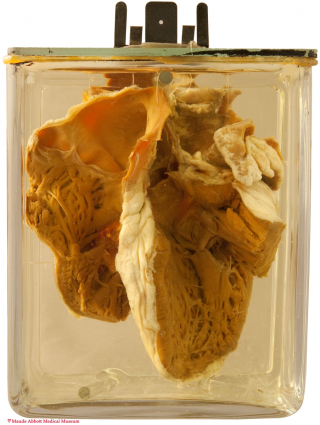
The specimen shows thickening (fibrosis) of the chordae tendineae to the anterior leaflet of the mitral valve (arrow, (left image)). An enlarged view of the tricuspid valve from the ventricular side (second image) shows fusion (short arrow) and thickening (long arrow) of the leaflets. A view from above (bottom image) shows the narrowed orifices of both mitral (M) and tricuspid (T) valves.


Comment
The specimen is the second case described in an 1877 report by Palmer Howard, Professor of Clinical Medicine at McGill when Osler was in Montreal. (Transactions of the Canada Medical Association 1877 I: 111 - 118). In this, he states that the heart came from the Museum and that there was no clinical record associated with it. A description of the specimen was given to him by Osler. It is not clear if the specimen came from one of Osler’s autopsies or had been donated to the Museum before he arrived in Montreal. Howard suggested that the cause of the stenosis was rheumatic fever.
![]() Canada Medical and Surgical Journal 1878, 6: 385 - 395
Canada Medical and Surgical Journal 1878, 6: 385 - 395