
Abbott Specimen 9
Specimen Card Nomenclature
Supernumerary aortic segment
International Classification of Diseases
Aortic valve cusp raphe
Atlas Illustration
None
Donor
Dr. Grunner
Date
1910
Age
Fifty years
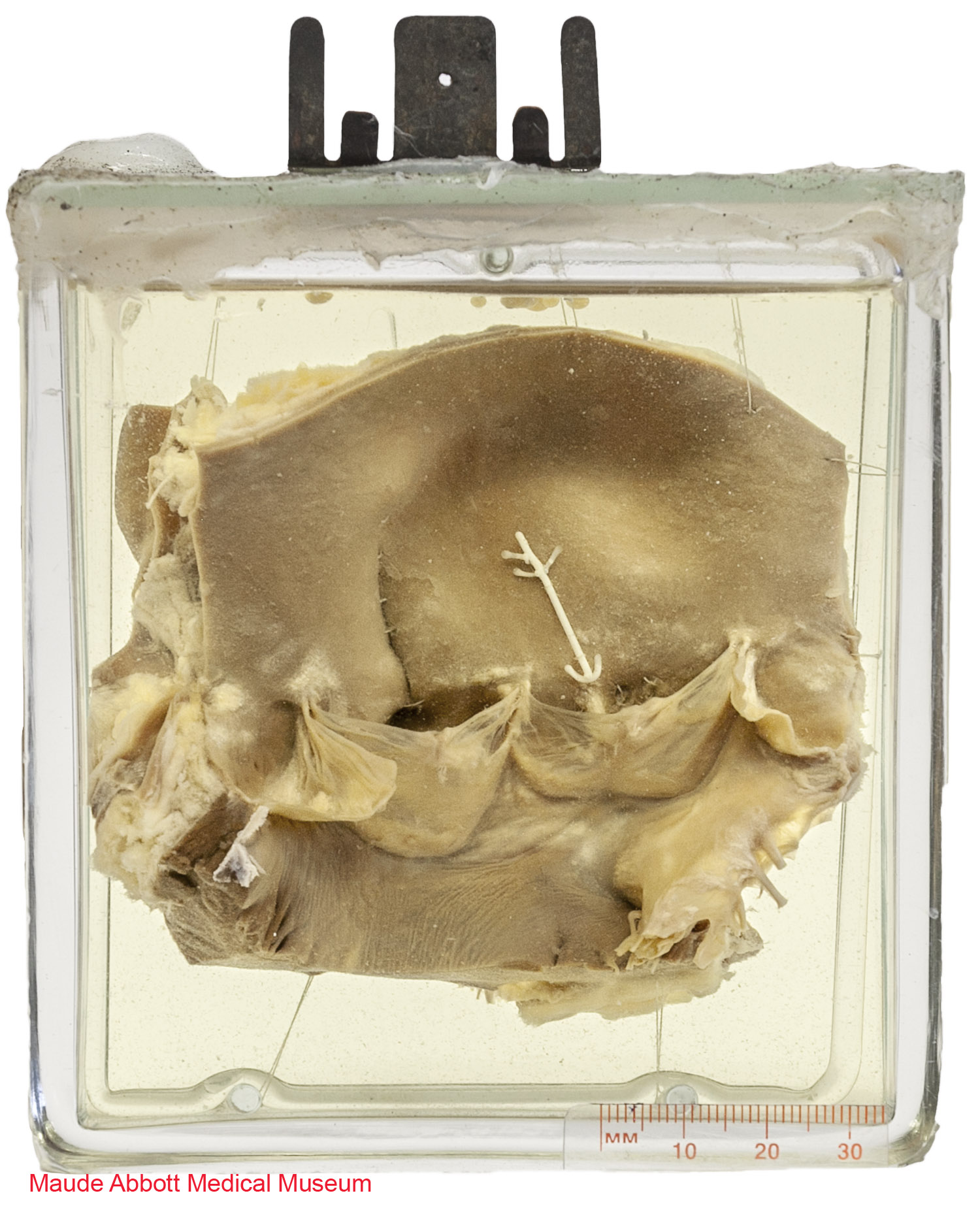
Description
The specimen shows an aortic valve with three leaflets (one cut in two during prosection). The leaflet on the right has a prominent fibrous band (raphe) extending from its base to the aortic surface (indicated by the arrow on specimen).
Comment
The aortic and pulmonary valves develop as swellings of the endocardial cushions on the inner aspect of the truncus arteriosus. When the four cushions fuse in the midline to divide the ventricular chambers, two additional swellings appear centrally. Continued growth is accompanied by excavation on the truncal side, resulting in the formation of sinuses between the presumptive leaflets and aorta/pulmonary artery wall. The precise mechanisms by which additional leaflets are formed or leaflets fail to develop are unclear. The presence of a fibrous raphe in an otherwise normal aortic valve (such as seen in this case) is rare and probably of no functional significance. Its cause is also unclear.
Figure 12:21. Langman, Jan. Medical Embryology: Human Development⎼Normal and Abnormal. Baltimore: The Williams & Wilkins Company, 2nd ed, 1969.
