Abbott Specimen 25
![]() Enlarge View
Enlarge View
Specimen Card Nomenclature
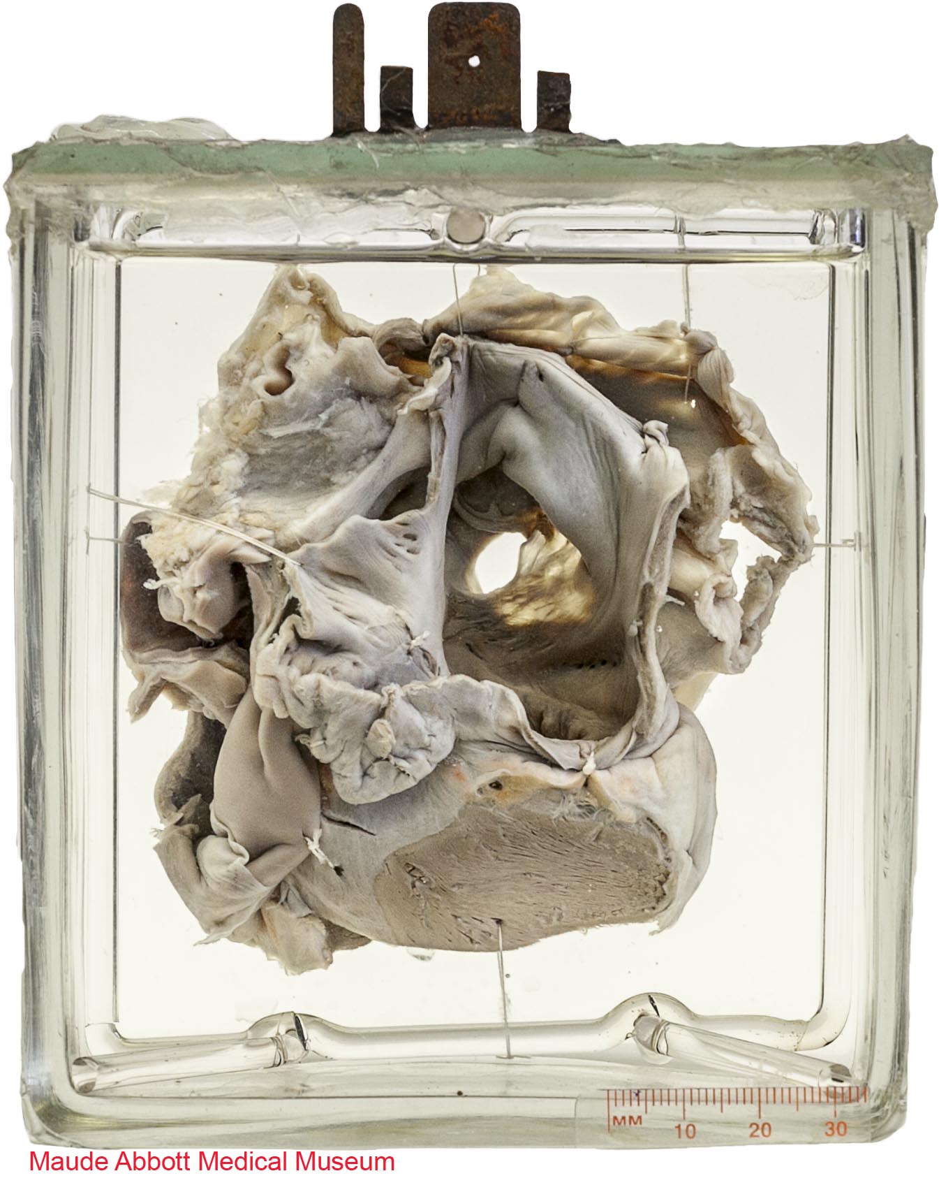
Heart of child with large patent foramen ovale and multiple defects of the inter-auricular septum
International Classification of Diseases
Atrial septal defect within oval fossa
Atlas Illustration
None
Donor
Dr. R. L. Ellis
Date
1905
Age
Unknown
Description
Front view shows the left atrium (A) with a 1 x 0.8 cm defect (arrow) in the fossa ovalis (F) of the interatrial septum
Comment
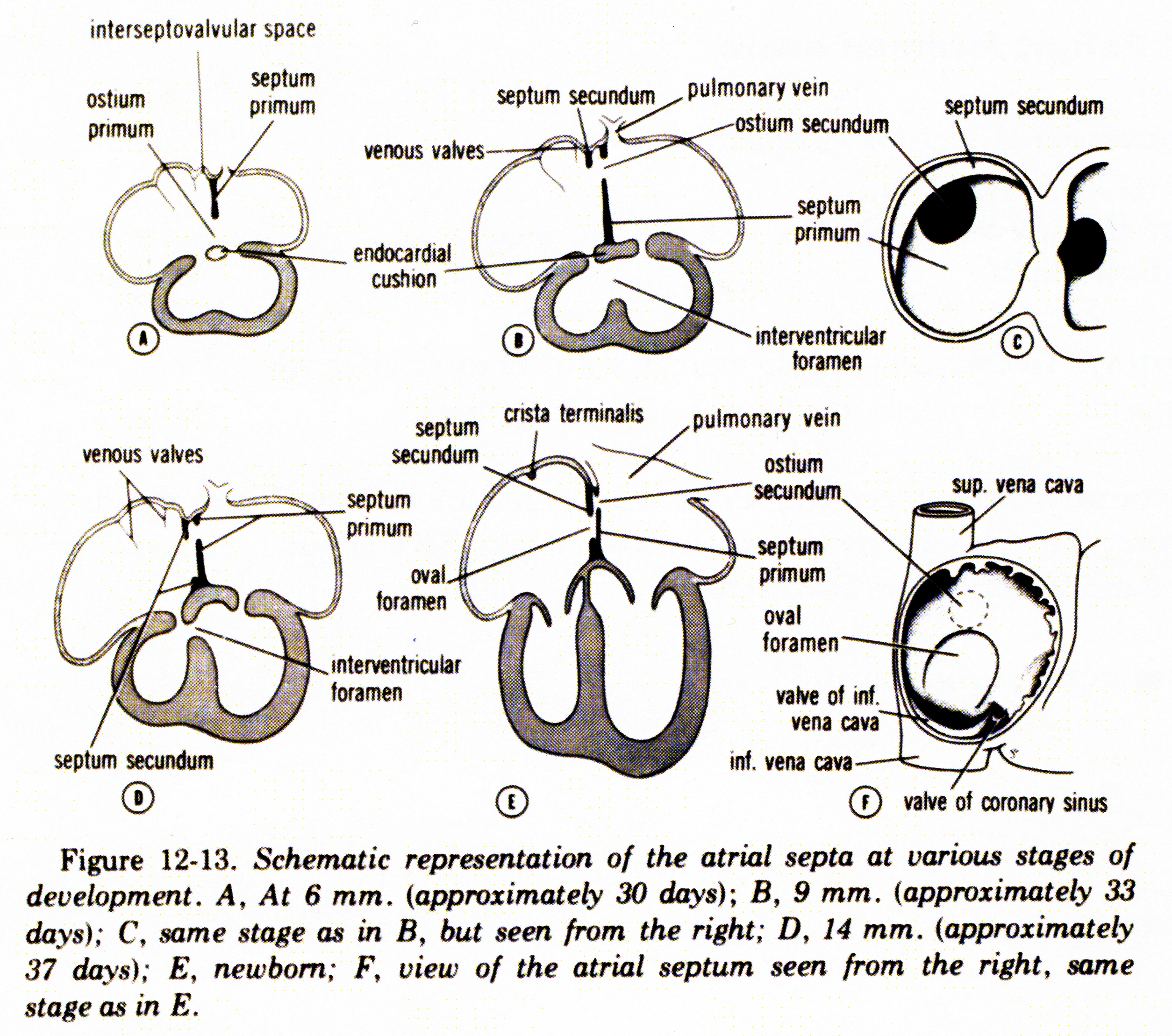
In early development, the atria are recognizable as two distinct bulges on either side of a common chamber. As these enlarge, a fold develops at the top and extends inferiorly (the septum primum), eventually fusing with the endocardial cushions at the ventricular junction. This process continually narrows the inter-atrial communication (termed the ostium primum). Normally, the latter closes completely; however, before this occurs, the septum primum develops perforations which coalesce to form a second communication (the ostium secundum).
As the atria continue to develop, a second tissue fold (the septum secundum) is produced to the right of septum primum. Normally, it does not develop over the entire septum primum, growing only to overlap the ostium secundum. The space between the two septa secundum at this place forms a continuity (the foramen ovale) that allows blood to flow from the right to the left atrium during fetal life. After birth, with increased pulmonary blood flow and elevation of left atrial pressure, the septum primum is pushed against the septum secundum, effectively closing the ostium secundum. Fusion of the septum primum and the septum secundum completely closes the foramen ovale in most individuals.
An ostium secundum defect such as seen in this specimen occurs when there is excessive resorption of the septum primum or incomplete development of the septum secundum. Although compatible with life (as demonstrated by the age of the patient from whom this specimen originated), the communication can be associated with several complications, including pulmonary hypertension and right heart failure (secondary to a left to right shunt), arrythmia (as a result of right atrial dilatation) and systemic (“paradoxical”) thromboembolism (resulting, for example, in stroke).
![]()
Figure 12:13. Langman, Jan. Medical Embryology: Human Development⎼Normal and Abnormal. Baltimore: The Williams & Wilkins Company, 2nd ed, 1969.