Mom Always Said, "Don't Rub Your Eyes. Good Advice for Eye Conditions, Especially Keratoconus"
What is Keratoconus?
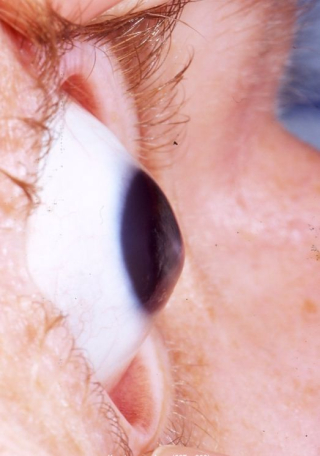
Keratoconus is a slowly progressive eye disease in which the normally round, dome shaped cornea (the clear outer front portion of the eye) thins and begins to bulge into a cone-like shape. This cone shape is irregular, bending light as it enters the eye, causing distorted vision. Since the cornea is responsible for refracting most of the light coming into your eye, developing an irregular cornea will cause blurred vision.
Keratoconus can occur in one or both eyes and often begins during a person's teens or early 20s. The condition occurs in approximately 1/1000 to 1/2000 people with varying degrees. Blurred vision caused by keratoconus (especially in its late stages) is different from blurred vision that is caused by other common refractive disorders such as myopia or hyperopia. In the later stages of keratoconus achieving satisfactory vision even with glasses or contact lenses can be difficult. Common refractive errors by comparison are easily correctable.
The "conical cornea" that is characteristic of keratoconus
Signs and Symptoms
To patients, early indications of keratoconus are usually a blurring and distortion of vision. In the early stages this may be barely noticeable, and only specific tests may uncover this condition. The need to frequently change glasses or contacts due to irregular shifts in astigmatism, changes in the curvature of the eye, or thickness of the cornea may be an indication that more specific tests, such as corneal topography, are required. Those with a progressive case will likely experience the effects gradually over several years. Occasionally, the thinning can progress more rapidly.
In the advanced stages of keratoconus, the patient may experience a sudden clouding of vision in one eye that clears over a period of weeks or months. This is called "acute hydrops" and is due to the sudden infusion of fluid into the stretched cornea. In advanced cases, superficial scars form at the apex of the corneal bulge resulting in more vision impairment.
Corneal Collagen Cross-Linking
Corneal Collagen Cross-Linking with Riboflavin (CXL, sometimes also abbreviated as C3R) is a minimally-invasive corneal treatment shown to slow or halt the progression of keratoconus. It does so by increasing the strength of corneal tissue. Undergoing CXL in the early stages may help stabilize vision. While the treatment will not correct vision or eliminate the need for glasses and/or contact lenses, it can help maintain the current level of vision and is meant to prevent vision from worsening. It can also help those that cannot wear contact lenses to more easily fit into them. The procedure is done in four steps.

1) The surface skin of the eye is gently polished using a rotary ‘epithelial’ brush.
2) Riboflavin (vitamin B2) drops are applied to the eye.
3) UV-A light is applied to the eye.
4) A bandage contact lens is put in place to assist the initial healing process.
How Corneal Collagen Cross-Linking Works
The procedure involves treating the eyes with riboflavin—vitamin B2—drops, and then exposing the cornea to 15-30 minutes of UV-A light. When combined with the riboflavin, it causes a reaction that increases the collagen bonds in the cornea which have been weakened by keratoconus. The treatment induces the natural anchors within the corneal fibers to link and prevent more bulging. Cross-linked corneal fibers actually occur naturally with aging. CXL speeds up that process and intensifies it.
Benefits of Corneal Collagen Cross-Linking
* It helps prevent further deterioration of vision
* Greater corneal rigidity, increased corneal resistance and biomechanical stability
* Prevention of keratoconus disease progression
* May defer the need for a corneal transplant procedure
* May reduce the myopia and astigmatism associated with keratoconus
* Enhanced contact lens wear
Prepared by Dr. Avi Wallerstein