
Table of Contents
Key words: maple, pigments, chlorophyll, carotenoids, anthocyanins, sucrose, osmosis, senescence, sap chemistry.
Abstract
This paper examines how the pigment chemistry of maple leaves and sap composition serve as molecular design solutions that enable the maple tree’s survival. As Autumn approaches, temperatures drop, and sunlight becomes scarce, chlorophyll is rendered ineffective for photosynthesis, marking the onset of leaf senescence. The maple tree has adapted to be resourceful; it recycles unnecessary chlorophyll molecules through a complex chemical process. Chlorophyll degradation occurs through a controlled pathway to dismantle and extract valuable nutrients, storing and recycling them. There are three main pigments responsible for the colors in maple leaves: Chlorophylls (A and B), Carotenoids, and Anthocyanins. As chlorophylls break down, carotenoids emerge while anthocyanins are newly synthesized, producing the yellow and red hues observed on maple leaves. These pigment transitions are governed by the hormones ethylene and abscisic acid. Through this system of processes, maple trees transform environmental and oxidative stress into a method of nutrient recovery and defense. In early spring, the maple tree’s sap enables nutrient transport through osmotic pressure-driven mechanisms. The high concentration of sucrose lowers the freezing point of water and creates osmotic gradients that maintain sap flow during freeze-thaw cycles, facilitating the regrowth and circulation of nutrients after dormancy. These chemical processes showcase the precision of adaptation. An understanding these processes provides valuable insight into potential biomimetic applications in materials science or biochemical engineering.
Introduction
The maple tree (Acer), a Canadian emblem and defining feature of forests across North America and Europe, is renowned for its resilience in colder climates and vibrant fall foliage, behind which lies a complex chemical system that enables it to endure harsh seasonal shifts. Chemical, alongside physical and other mechanisms unique to the maple tree give it unique adaptive qualities that act as and inspire design solutions to problems posed by its geographical distribution. (see Maple Physics for an extensive introduction)
Leaf Coloration
The spread of vibrant colors in Canada’s boreal forest is created by deciduous trees, including the Maple tree. This colorful mosaic occurs during autumn when environmental stressors, such as the harsh temperatures of the northern hemisphere and reduced light exposure, begin to greatly impact the ecosystem (Schaberg et al., 2008). The different compositional elements of the leaf, specifically the various pigments and their fluctuating quantities, are responsible for the vivid colors of foliage. Indeed, chlorophylls, carotenoids and anthocyanins are the three main pigments that determine the display of colors in a leaf, which vary from green (Fig.1 c) to yellow (Fig. 1 b), to even a brilliant red (Fig.1 a). These pigments can be classed according to their moment of formation during the maple leaf’s lifespan. In fact, both chlorophylls, that express green, and carotenoids, responsible for yellow, are developed during the beginning of the leaf’s growth period. Furthermore, anthocyanins, which generate the reddish hue of maple leaves during autumn, are synthesized near the end of the deciduous cycle (Schaberg et al., 2008).

Fig. 1. Different maple leaf colorations. A) red leaves, b) yellow leaves and c) green leaves.
Pigments and Their Role in Coloration
1. Pigments Responsible for Green: Chlorophylls
Maple leaves, like all other vascular plants, are composed of plant cells that undergo photosynthetic processes in the interest of transforming light energy into chemical energy (Fowler et al., 2024). Indeed, incident light energy stimulates light-dependent reactions within chloroplasts, which are the photosynthetic apparatuses of a plant cell. Chloroplasts are composed of grana, which are stacks of small green disks that are called thylakoids (Fowler et al., 2024). The light-dependent reactions of the photosynthetic process occur in the thylakoid membrane, where chlorophylls, such as chlorophyll a and chlorophyll b (Fig.2), absorb solar radiation and transform it into chemical energy (Fowler et al., 2024).

Fig. 2. Chemical formulas and structures of chlorophylls. a) chlorophyll a. b) chlorophyll b, modified from Scifinder software.
Light absorption of different wavelengths occurs in the light sensitive complexes found in the thylakoid membrane (PSI/PSII) and is responsible for the green coloration of leaves. Indeed, the photosystem II (PSII) and the photosystem I (PSI) both use chlorophyll pigments to absorb blue wavelengths (400-500 nm) and red wavelengths (600-700 nm) (Liu & van Iersel, 2021). However, green wavelengths (500-600 nm) are poorly absorbed by these photosynthetic complexes, hence the green color of leaves (Liu & van Iersel, 2021).
2. Pigments Responsible for Yellow: Carotenoids
During normal photosynthetic processes, chlorophylls are continuously broken down and replaced with new synthesized ones (Şahin & Onay, 2023). However, when the leaf begins its deterioration phase, the production of chlorophyll is suppressed unmasking the presence of other pigments in the leaf, such as carotenoids like carotene-b (Fig.3 ) (Şahin & Onay, 2023).

Fig. 3. Chemical formula and structure of β-carotene, modified from Burton & Ingold (1984).
Carotenoid pigments are first synthesized in the chloroplast during the leaf’s growing period. These molecules are primarily responsible for the yellow and orange coloring of leaves but they are normally overpowered by the presence of chlorophylls, which are stronger pigments(Şahin & Onay, 2023). However, with the arrival of autumn, extreme conditions, such as colder temperatures and limited light, induce the degradation of the structural components of leaves, including pigments (Moy et al., 2015). Moreover, the deterioration rate of chlorophylls is much faster than that of carotenoid molecules (Moy et al., 2015). Thus, as the chlorophylls begin to dismantle, carotenoids are revealed, expressing a yellowish hue in the leaves.
3. Pigments Responsible for Red: Anthocyanins
The biosynthesis of anthocyanin pigments, responsible for the reddish leaves of Fall, was previously thought to have been caused by structural deterioration with age (Schaberg et al., 2008). However, this hypothesis was proven to be faulty following the observation of increased anthocyanin production in leaves that had been exposed to external stressors. Environmental factors, such as low temperatures, UV-B radiation, osmotic stress and lack of nutrients, have been shown to enhance the biosynthesis of anthocyanin pigments (Schaberg et al., 2008). Therefore, it is possible to conclude that anthocyanin production is prompted in the interest of countering stress-related harm.
During autumn, the leaf begins to degrade, starting with the deactivation of photosynthesis and the photodamage of PSII, also known as photoinhibition (Moustaka & Moustakas, 2025). Thus, light absorbing structures such as chlorophyll and photosystems are overexcited and cannot safely dissipate solar energy, leading to excessive energy in the plant cell. These extreme light intensities cause oxidative stress and can generate the production of harmful reactive oxygen species (ROS) (Moustaka & Moustakas, 2025). Once fabricated, the ROS can modify the structural composition and function of essential biomolecules, thereby significantly damaging and potentially killing the plant cell. (Sachdev et al., 2023). Furthermore, anthocyanins play a key role in decreasing photooxidative damage. In fact, these pigments are often compared to sunscreen, seeing that they reduce the amount of light captured by leaves (Schaberg et al., 2008). In addition, anthocyanins can reduce harm caused by ROS due to their antioxidant qualities, which is their ability to inhibit oxidation. Thus, anthocyanin, unlike other leaf pigments, are synthesized during the later stages of a leaf’s lifespan to reduce photooxidative damage (Schaberg et al., 2008).
4. Change in Color of Leaves Between Years
Maple trees' color vibrancy varies yearly. During some years, maple trees exhibit strong red colors for an extended period, whereas during other years, the colors appear duller and do not persist for long. This variability can be attributed to different factors: exposure to sun irradiation, temperature, water and nutrient shortage, herbivory or disease (Lev‐Yadun, 2022). The time of senescence also plays a role in the development of the red color, as red maple trees with early senescing leaves have higher anthocyanin content, which is responsible for the red color as aforementioned, compared to late senescing leaves. Early senescing can be explained with multiple environmental stressors, such as nitrogen limitation, cool daytime temperatures, canopy openness (i.e. the overlap of tree crowns) and soil pH (Anderson & Ryser, 2015). Prolonged and frequent rain and high humidity during the summer lead to the proliferation of foliar fungal diseases, which can impact productivity and cause brown spots on leaves. Other meteorological factors are at play, as cloudy weathers can reduce the irradiance required by leaves to anthocyanin and intense red hues (Caduto, 2024).
Degeneration of Chlorophylls
1. Leaf Senescence
Leaf senescence is characterized by the deterioration of the structural composition of foliage (Thakur et al., 2016). This is the last period of the leaf’s lifespan during which anabolic reactions, specifically photosynthesis, are replaced by catabolic reactions that break down the different macromolecules composing the structures of the plant cell. During this decomposition, nutrients are produced and remobilized by the maple tree for survival during dormancy and for regrowth. The gradual loss of function and the deterioration of cells during leaf senescence eventually cause the death of the maple leaves, characterized by their detachment from the tree (Thakur et al., 2016). Moreover, the degradation of green chlorophyll pigments is a crucial occurrence during senescence. This breakdown is generated by various external stressors and can be described by a series of events that will be explained below.
2. Initiation of Senescence by Hormonal Signalling
Senescence is initiated by seasonal changes. Indeed, in autumn, nights begin to get longer while days are shorter, and temperatures begin to drop. These factors serve as a signal of winter’s arrival and induce hormonal changes within the leaves. In fact, the aging of leaves is regulated by a network of phytohormones that either promote senescence (e.g. ethylene, jasmonic acid, abscisic acid and salicylic acid) or subdue it (e.g. cytokinin, auxin, gibberellic acid, nitric oxide, and polyamines) (Zhang & Zhou, 2013). The balance of these hormones, which is a determining factor of senescence occurrence, is modified by insufficient light and extreme temperatures.
Ethylene is considered the hormone that plays the most crucial role in the promotion of leaf senescence (Zhang & Zhou, 2013). The hormonal regulation of ethylene involves a MAP kinase (MAPK) signaling cascade, which is a network of protein kinases that regulate cellular processes (McManus, 2012). Indeed, ethylene production can be described by the Yang cycle, which involves two enzymatic steps (McManus, 2012). First the production of 1-aminocyclopropane-1-carboxylic acid (ACC) is promoted by the ACC synthase enzyme (ACS):
ACC Synthase (ACS)
S-Adenosylmethionine (SAM) → 1-aminocyclopropane-1-carboxylic acid (ACC)
Then, ACC is converted into ethylene with the help of the ACC oxidase enzyme (ACO):
ACC oxidase (ACO)
ACC → Ethylene + CO2 + HCN
Furthermore, ACS and ACO expression are promoted by environmental factors (e.g. darkness and cold) leading to the upregulation of ethylene during leaf senescence (McManus, 2012).
Following this hormonal increase, several membrane receptors found in the endoplasmic reticulum (ER) perceive the ethylene signaling. Indeed, in the presence of ethylene, these receptors bind the hormone and change its conformation, which inactivates Constitutive Triple Response 1 (CTR1), a protein kinase that inhibits the signaling of ethylene insensitive 2 (EIN2) (McManus, 2012). EIN2 is a membrane protein that transmits the ethylene signaling to the nucleus, thereby stabilizing EIN3 and EIL1, leading to their accumulation in the nucleus. Ethylene-insensitive 3 (EIN3) and its homolog (EIL1) are transcriptional activators that regulate ethylene signaling (McManus, 2012). Once they have been stabilized and accumulated, EIN3 and EIL1 create a positive feedback loop by directly increasing ethylene biosynthetic enzymes (McManus, 2012). The positive feedback loop of ethylene production allows the tree to quickly and irreversibly transition from being photosynthetically active to being dormant, preparing it for survival during winter.
Moreover, EIN3 and EIL1 can also play a key role in the degradation phase of leaves by activating Senescence-Associated Genes (SAGs) and Chlorophyll Catabolic Genes (CCGs) (McManus, 2012). In fact, once activated, they can control and reprogram ethylene-responsive genes (ERGs), allowing them to initiate certain processes, such as chlorophyll degradation, protein catabolism, and nutrient remobilization.
Many other hormones, including abscisic acid (ABA), jasmonic acid (JA) and salicylic acid (SA), act as senescence enhancers (Zhang & Zhou, 2013). Despite having different mechanisms, they all work towards the same goal, which is to induce the degradation of leaves. Furthermore, there also exists several hormones that delay senescence, such as cytokinin, auxin, gibberellic acid, nitric oxide, and polyamines (Zhang & Zhou, 2013). With the arrival of autumn and its harsh conditions, these senescence suppressor hormones also undergo specific mechanisms, that result in a decrease in their production. Indeed, each suppressor hormone has a regulatory pathway associated with senescence, such as biosynthetic, transport and signalling pathways. Overall, the hormonal shift, caused by a drop in temperature and limited access to light, reduces photosynthetic activity and promotes degradation of chlorophyll and of biomolecules, leading to nutrient recycling.
3. Deactivation of Photosynthesis
Following the shift in hormonal regulation, the metabolic activity of the leaf is reprogrammed, altering from anabolic, which involves synthetic processes, to catabolic, which involves the breakdown of larger molecules into smaller ones (Zhang & Zhou, 2013). Indeed, when the leaf begins its deterioration phase, senescence promoting hormones suppress photosynthetic genes, leading to the dismantling of chloroplasts which are responsible for photosynthesis (Zhang & Zhou, 2013). On top of this transition, the deactivation of photosynthesis is further influenced by the insufficient light of shorter autumn days (Hu et al., 2025). Indeed, photosynthesis decreases according to the amount of light available for the chlorophyll pigments to absorb (Hu et al., 2025).
4. Disassembly of Photosystems
After loss of photosynthetic activity, the photosystems (PSI and PSII) still absorb solar radiation and excite electrons. However, these electrons can no longer be used to generate chemical energy and, having nowhere to go, can cause oxidative damage (Moy et al., 2015). Indeed, the deactivation of photosynthesis causes over-reduction in the electron transport chain (ETC), promoting the leakage of electrons to oxygen and forming ROS. Thus, PSI/PSII are light-sensitive complexes that begin to disassemble during senescence in the interest of limiting excess electrons and reducing the production of ROS (Moy et al., 2015). During the dismantling of these photosynthetic complexes, the chlorophylls, that were tightly bound to the photosystems by proteins, are released (Moy et al., 2015). Moreover, these pigment molecules are also subjected to degradation during senescence (Tanaka & Ito, 2025).
5. Chlorophyl Degradation Pathway (PAO Pathway)
The catabolic reaction that occurs in chlorophylls can be described by the chlorophyll degradation pathway, also called the PAO/Phyllobilin pathway (Tanaka & Ito, 2025). This highly regulated enzymatic process occurs during leaf senescence and allows the tree to safely decompose the green chlorophyll pigment and recycle its nutrients. This pathway describes the conversion of chlorophylls into colorless phyllobilins that are stored in the vacuole(Tanaka & Ito, 2025). The first step towards chlorophyll breakdown is the conversion of chlorophyll b to 7-hydroxymethyl chlorophyll a using chlorophyll b reductase (NYC1 and NOL) (Fig.4). This initial transition is essential since most enzymes can only act on chlorophyll a and its derivatives. During the second step, 7-hydroxymethyl chlorophyll a reductase (HCAR) is the enzyme that promotes the formation of chlorophyll a from 7-hydroxymethyl chlorophyll a (Fig.4). From this point both chlorophylls follow the same degradation pathway. The third step in this catabolic process involves the removal of the central magnesium ion (Mg2+) from chlorophyll a using the Mg-dechelatase, Stay-Green (SGR), and resulting in the formation of pheophytin a (Fig.4). This part of the pathway is responsible for the partial deactivation of chlorophyll’s light absorbing center. The dismantling of the light absorbing center limits the generation of ROS that can be harmful to the cell. Following this step is dephytylation, the removal of the phytyl tail of pheophytin a, which is catalyzed by pheophytinase (PPH) and yields pheophorbide a (Fig.4). The fifth step, the ring cleavage, is the most crucial part of the PAO pathway. This step is completed using the pheophorbide a oxygenase (PAO) enzyme, a non-heme iron monooxygenase, to open the porphyrin ring between C5 (of ring A) and C10 (of ring B). Indeed, the methine bridge(-CH=) connecting the two upper pyrrole rings is broken using ferredoxin and O2 for oxidative cleavage. This ring-opening step, converting pheophorbide a to red chlorophyll catabolite (RCC) (Fig.4), is essential because it completely cancels out chlorophyll’s light-absorption capabilities. Step 6 involves a further reduction of RCC using RCC reductase (RCCR/ACD2) that manipulates electrons from ferredoxin to breakdown RCC, forming primary fluorescent chlorophyll catabolite (pFCC) (Fig.4). Following this step in the pathway, the catabolites are exported from the cytosine and undergo a series of slight modifications near the chloroplast ER that render them more soluble, facilitating vacuole storage. Finally, the seventh and last major step in the PAO pathway is the isomerization of catabolites that occurs following their transportation to the vacuole. In fact, once inside the vacuole, pFCC is converted to nonfluorescent chlorophyll catabolite (NCC) (Fig.4), making the final molecule stable and entirely colourless (Tanaka & Ito, 2025).

Fig. 4. Degradation of chlorophyll pigments using the PAO pathway (Tanaka & Ito, 2025).
6. Nutrient Remobilization
Leaf senescence is accompanied by nutrient recycling that occurs over the course of the degradation of cellular structures and their macromolecules (nucleic acids, proteins, carbohydrates and lipids) (Avila-Ospina et al., 2014). Indeed, this process involves reclaiming essential nutrients, including nitrogen, phosphorus and magnesium, from dead tissue before the leaf sheds (Maillard et al., 2015). These nutrients are remobilized, transported through the phloem, a plant vascular tissue, and stored in tissues in the branches and roots of the tree (Guo et al., 2021). They ensure the tree’s survival through winter and promote leaf growth in the spring. After senescence has been initiated, the deactivation and dismantling of photosynthetic apparatuses lead to an accumulation of nutrients. Indeed, with a decrease in photosynthesis, chloroplasts and its internal structures begin to degrade, leaving behind macromolecules from which nutrients are recycled (Guo et al., 2021). In fact, nitrogen is often recovered as amino acids, nitrate or nitrogen and is the result of protein breakdown (Hortensteiner & Feller, 2002). For example, proteases are used to degrade RuBisCo, an important protein in leaf composition, resulting in the production of amino acids (Hortensteiner & Feller, 2002). Moreover, phosphorus is recycled by nucleases and phosphatases that degrade ATP, nucleic acids (DNA and RNA) and phospholipids (Stigter & Plaxton, 2015). Finally, magnesium is retrieved during the third step of the PAO pathway in chlorophyll degradation, when chlorophyll a is converted to pheophytin a using Mg-dechelatase (SGR) (Tanaka & Ito, 2025).
Chemical Composition of Sap
Carbohydrate Storage
Maple trees thrive in environments with long and cold winter seasons. Due to harsh environments where food resources can be scarce and sunlight dim, maples adapt by developing a storage system for carbohydrates and other essential nutrients to recover after the dormant stage. They produce carbohydrates through photosynthesis during the summer and store them as energy reserves, which can be mobilized at the beginning of spring through cell respiration to transition into the growing stage. In fact, during sap exudation, concentrations of CO2 are higher than O2 content compared to ambient air, showing respiratory activity peaks during this period, and carbohydrate reserves are being used by the tree to grow back its leaves to start photosynthesis again (Perkins & van den Berg, 2009). In addition, sap contains minerals originating from the soil, organic components, and amino acids in their roots, which are also related to the tree’s metabolic activity. Once temperatures start to rise with the arrival of spring, the sap moves upwards through numerous interdependent processes, like cohesive-tension forces, osmotic forces, and pressure fluctuations that happen because of spring temperature fluctuations. The chemical composition of sap also contributes to sap flow (Zimmermann et al., 2002).
Studies throughout the decade show that over two hundred natural and process-induced compounds have been detected in maple sap. Among those, the main compounds are sugars, water, organic acids, amino acids, proteins, pyrazines, phenolic compounds, and vitamins (Table 1). Detailed insight into chemicals contained in sap is the result of gas chromatography, mass spectrometry, and nuclear magnetic resonance. Additionally, metal compounds in fluctuating concentrations have been found (Mostly calcium and potassium) (Ball, 2007). These fluctuations are hypothesized to be the result of climatic and soil compositions depending on the year, and possible sap contamination during extractions (Mohammed et al., 2022).
Table 1. Organic components found in Maple sap (Ball, 2007)
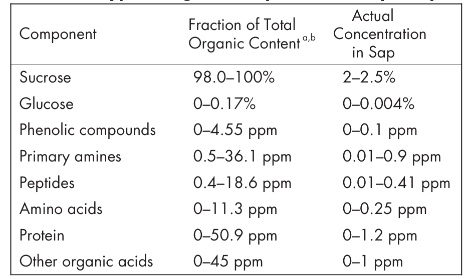
Maple sap is mainly composed of water, comprising between 95 and 99%, and sugar, constituting 1 to 5% of sap, for sugar, black, and red Maple specifically (Acer Saccharum, Acer Nigrum, and Acer Rubrum respectively), with amino acids, organic acids, proteins, minerals and phenols in lower concentrations (Perkins & van den Berg, 2009).
Growing season is a period where growth theoretically takes place. That is, between the day of the last spring freeze and the first autumn freeze (Linderholm, 2006).
Soluble sugars like saccharose peak in production during the winter and near spring, while starch, the main carbohydrate reserve, is low throughout summer, but accumulates gradually in the wood and twigs until the end of the growth season. This accumulation of sugar reserves serves as energy storage for the leafless period. Soluble sugar grows in quantity during winter and spring. Saccharose, also known as sucrose, is the main soluble sugar found in the xylem (vascular tissue). As for other sugar molecules, only minor quantities of glucose and fructose are present. Even less stachyose, raffinose, and xylose are detected (Perkins & van den Berg, 2009).
First, the significant presence of sugars in maple sap serves as an energy reserve during the early spring when no photosynthesis occurs. It allows sufficient energy production for bud break and leaf expansion. Soluble sugars extracted originate from carbon reserves in adjacent parenchyma cells, or from hydrolyzed starch reserves in the roots (Muhr et al., 2016). Maples tend to store carbohydrate gains from several previous growing seasons and mobilize the energy source over the years following production, with both ‘‘old’’ and ‘‘new’’ sugars released into sap. In fact, experiments on maple sugar reserves using the bomb-spike radiocarbon technique show that accumulated starch can be a couple of years old. In the 1960s, nuclear testing led to spikes in radioactivity, resulting in peak levels of carbon-14 and carbon-13 isotopes. Consequently, radiocarbon testing is used to trace carbon isotopes, mainly carbon-14, in non-structural carbon, including sugar in sap, to indicate for how long it is stored. Measurements of tree carbon-14 signatures show that non-structural carbon is usually stored for three to five years before being used (Fig. 5). Storing starch in intervals of years prepares the tree for unique harsh conditions or strong masting events, when more energy is necessary than the reserve pool of a single growing season (Muhr et al., 2016). Therefore, maple trees release a mixture of recently stored carbohydrates and old reserves in sap for metabolic and growth functions.

Fig. 5. Tree carbon-14 signature (Δ14C) of two populations of maple trees. Δ14C values in the range of 30% to 40% indicate that the average storage period of non-structural carbon is around three to five years old (Muhr et al.,2016).
Specifically, sucrose is the carbohydrate precursor for cellulose synthesis. It is a disaccharide formed through an alpha glycosidic bond between glucose and fructose. Once synthesized, sucrose is transported in maple sap to the tree cells for cellulose formation. Within these cells, sucrose is broken down into its monosaccharide constituents, and the glucose is polymerized via beta glycosidic bonds to form cellulose, while fructose is used for other metabolic functions (Fig. 6). The decomposition of saccharose into its two monosaccharide constituents during the cellulose formation explains the trace presence of fructose and glucose as secondary sugars to saccharose present in sap (Ball, 2007).

Fig. 6. General Structure of sucrose and cellulose (Ball, 2007).
Contribution of Sugar Content in Sap flow
Second, the high sugar concentration contributes to the sap flow, allowing better transportation of the liquid upwards. Maple sap circulation, often called exudation, occurs due to multiple mechanisms. Among the main ones is temperature fluctuation. At the beginning of spring, drastic changes in temperatures between the day and the night occur (shifting roughly between 10°C to -10°C (West et al., 2023), causing variations in pressure that lead the sap to circulate. This phenomenon is also known as the freeze-thaw process. The tree trunk of maples is composed of water (and sap) circuits that allow longitudinal movement as a main transport of sap. However, sap movement also occurs in radial and circumferential directions (Fig. 7). In maples, the peripheral area of the trunk is called sapwood, which constitutes the area in which sap flow occurs. Sapwood contains living and dead wood fiber cells collectively composing the xylem, and they are filled with gas (Tyree, 1984) as shown in figure 7, before exudation begins (a). When temperatures begin to decrease, water contained in the fiber cell gradually freezes (b), forming a frost like layer inside of the cell wall (Timothy D Perkins & Abby K van den Berg, 2009). As a result of the lower temperature and vapor pressure of ice, the gas pressure drops, causing the water in the xylem vessels to be drawn into the fiber cells and freeze (c). The entry and expansion of water when it solidifies causes the pressure to increase, along with the rise of temperatures that warms the tree (d) (Robinson et al., 2024). As a result, sap is pulled towards the top of the tree during the night and freezes. Once the temperature increases above freezing, the sap flows down the tree by gravity and pressure increases.
Xylem, along with the phloem constitute the vascular system in most land plants and appeared soon in plant evolution. It is the system that controls sap flow.
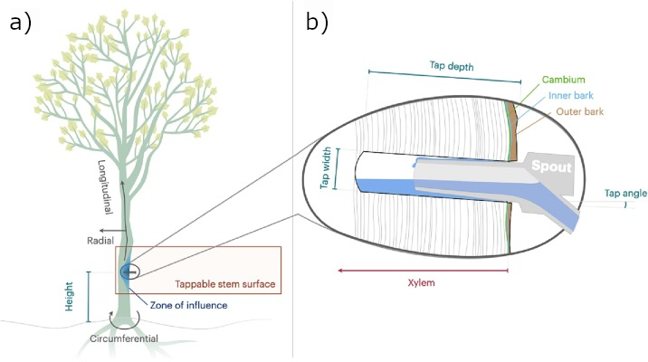
Fig. 7. Sap transportation and tapping. (a)Transport of sap happens longitudinally, radially, or circumferentially (gray arrows), although it primarily occurs in the longitudinal axis. (b) Close-up of tap hole with spout illustrating the tap hole depth, which is the location of the xylem, the outer portion of the stem (Adapted from Rademacher et al., 2023).

Fig. 8. The freeze-thaw mechanism in four steps, showing the pressure changes in a xylem fiber from the initial state (a) to the last (d) (Robinson et al., 2024)
When the pressure inside the fibers is high, meaning it is above or equal to atmospheric pressure (d), the gas pressure inside the cell fibers increases beyond air pressure on the trees' surface, causing the gas to dissolve in the liquid. The presence of water causes gas pressure to increase even more because of surface tension acting on the air bubbles, thus accentuating the gas diffusion. Theoretically, no air is left within the fiber cells, preventing the alternating pressures leading to exudation. This is an additional component, preventing gas diffusion and thus partaking in sap flow.
Recent discoveries stipulate that high saccharose content in sap is correlated with sap yield. Later, after this observation, researchers identified the physiological process responsible for this relationship to be the osmosis forces that add additional pressure on fiber cells, hence preventing gas from diffusing. As a result, the high sugar content not only serves as an energy reserve, but it also contributes to the sap’s motion against gravity. It was hypothesized (as modelized in Fig. 9) that fiber cells and vessel cells are separated by a semi-permeable barrier, allowing the free movement of water, but restricts the movement of large molecules such as sucrose, the osmolyte in this context. The osmotic barrier stops the diffusion of saccharose that accumulates in the xylem during the winter season. Therefore, the high concentration gradient of saccharose between the fiber cell and the vascular cell drives part of the water in the fiber cell to penetrate the vessel. The osmotic pressure resulting from the movement of water decreases the pressure inside fiber cells. Consequently, the gas bubbles do not dissolve and keep the pressure inside fiber cells to assure pressure variations, and therefore sap flow between the roots and the canopy. An experiment run by Johnson (1987) (Johnson et al., 1987) confirmed that high saccharose content facilitated exudation. Tree twigs were made to undergo freeze-thaw cycles to study the absorption and exudation of sap. Twigs perfused with 2% saccharose solution exudated during the thaw phase, while water-perfused twigs did not (Robinson et al, 2024). It is worth mentioning that sucrose might not be the only molecule causing osmotic pressure. Multiple solutes with high molecular weight are unable to cross the semi-permeable barrier. Thus, each component contributes relatively to osmotic pressure. However, the dominating presence of saccharose makes it the main actor in this mechanism.

Fig. 9. Simplified model of sucrose involvement in sap exudation: (a) When semi-permeable barrier is absent, there is free crossing of large molecules from the vessel into the fiber cell, the liquid pressure in the fiber Pf is equal to the liquid pressure is the vessel Pv. The gas pressure Pg is thus the sum of the liquid pressure and the water surface tension ∆Pst: Pg= Pf + ∆Pst. In this case, the gas pressure is beyond atmospheric pressure (Pg>Patm) which causes the diffusion of gas, preventing the pressure fluctuation process from happening. The model (b) illustrates the separation of both cell types by a semi-permeable membrane that only allows the crossing of water. The osmotic pressure (∆Pos) caused by concentration gradient differences causes the liquid pressure Pf to decrease, which implies a lower gas pressure Pg, thus preventing the diffusion of gas (Pg ≤Patm) (Robinson et al., 2024).
The osmotic pressure model was developed following experimental observations showing that sap flow increases with increasing sucrose concentration, indicating a positive correlation between sap yield and sugar content in sap. Among these experimental works, Johnson (1987) studied the response in sap flow of excised stems when perfused with either water or sucrose solutions. Stem segments free of lateral branches of sugar maple seedlings were collected, cut in the early morning, wrapped in damp paper and aluminum foil to keep them frozen. Other samples were collected in nonfreezing conditions (maintained at 4°C). The branches were kept in insulated boxes with pressure regulation, where they could be perfused with different solutions (in this case, water and sucrose). For reproducibility and uniformity of the experiments, freezing cycles were controlled, and the branches were perfused with a 10 mM NaCl solution to equilibrate overnight. Then, the stems were supercooled to -3°C until temperature and solution flow equilibration were reached. The freezing of the stem was induced, and after a period lasting between three to four hours, the stem was warmed for the same approximate amount of time at +3°C until equilibrium was reached. Then, the stem was perfused with 100-200 ml of a test solution (either water or 2% sucrose solution). Lastly, they measured the maple stem response to the freeze-thaw cycle. The results showed that, as the concentration of saccharose in sap increased, with the maximum value corresponding to the maximum content found in sap (5%), sap flow also increased (Fig. 10). From this observation, no mechanism responsible for this increase was identified (Johnson et al., 1987).

Fig. 10. Johnson’s results lead to the conclusion that sap flow increases with higher sucrose concentration in vessel sap, for the interval of saccharose content found in maple (up to 5%) (Johnson and al., 1987).
However, a study (Robinson et al., 2024) later further described the relationship between sap flow and sap sugar content. In fact, it was first highlighted that the freeze-thaw theory was incomplete, and the additional component contributing to the functioning of the freeze-thaw process was the osmotic effect of sucrose. The use of microCT allowed a better study of the phenomenon with imaging. The xylem was perfused with water and sucrose solutions at high resolution and without sectioning. Final observations led to conclude that an osmotic barrier was indeed present, but the saccharose effect on sap flow is more complex than initially modelized (Fig. 9), and additional parameters need consideration.
Finally, carbohydrate storage and sap flow of maple trees allow access to sufficient energy during the leafless period. In fact, carbohydrates are stored through several growing seasons and are readily available for cellulose synthesis no matter how energy-depriving environmental conditions can be. Furthermore, although some maple trees tend to produce sweeter sap, the selective factors are mostly misunderstood. As discussed, experiments show that maples that have higher sap yields tend to have higher sap saccharose concentration. However, increased sugar content has yet to be proven as a design solution to maximise sap flow, but is directly correlated to high sap yield, making it a plausible candidate (Rademacher et al., 2023). Thus, sugar concentration in sap is correlated with flow and contributes to osmotic pressure, which enhances sap yields, with other mechanisms, such as the freeze-thaw cycles in xylem.
Conclusion
Maple trees evolved to survive cold climates where the environment is harsh, and resources can be scarce in the winter by developing biochemical design solutions. First, maple undergoes leaf senescence and switches from anabolic to catabolic reactions during winter where days are shorter and the light available for photosynthesis is reduced. Ethylene, one of the major regulatory hormones for senescence, is produced as a result of darkness and cold. Following this hormonal shift, photosynthetic activity is reduced and chlorophylls are degraded. Anthocyanins are produced to counteract oxidative damage, as photosystems are disassembled, meaning the absorbed energy is not dissipated and causes damage to the cell. Anthocyanins and carotenes which take longer to be degraded lead to the yellow-red color of the leaf. Chlorophyll degradation undergoes a carefully orchestrated degradation process known as the PAO pathway. This allows the last product to stabilize and ensure recycling of the nutrients, which are remobilized in the tissue of branches or roots of the tree as the leaves die. Maple trees then enter a dormant state during the winter and rely on those resources. Maple sap is also crucial for the storage of carbohydrates during the winters. It has high sugar content, both soluble sugars and starches. Soluble sugars are used when photosynthetic reactions are halted. Starches are formed from the carbohydrate gain during the growing seasons and stored for multiple years to be used for unique harsh environments where more energy is needed. They can then be hydrolyzed into soluble sugars. The high sugar content in sap is crucial for sap transport as it lowers the freezing temperature of the sap, contributing to better pressure fluctuations in the freeze-thaw process and leading to better sap transportation. This pressure cycle occurs when nighttime freezing causes water to freeze, creating a small layer of frost and reducing the pressure, leading to the suction of sap towards the top of the tree. Thawing during the day leads to gas expansion and pressure increase, pushing the sap back down. The sugar in sap also leads to osmosis forces that add additional pressure on fiber cells and prevents gas from diffusing, assuring pressure variations and sap flow.
References
References
Anderson, R., & Ryser, P. (2015). Early autumn senescence in red maple (Acer rubrum L.) is associated with high leaf anthocyanin content. Plants, 4(3), 505-522. https://doi.org/10.3390/plants4030505
Avila-Ospina, L., Moison, M., Yoshimoto, K., & Masclaux-Daubresse, C. (2014). Autophagy, plant senescence, and nutrient recycling. J Exp Bot, 65(14), 3799-3811. https://doi.org/10.1093/jxb/eru039
Ball, D. W. (2007). The chemical composition of maple syrup. Journal of Chemical Education, 84(10), 1647-U1646. https://doi.org/10.1021/ed084p1647
Burton, G. W., & Ingold, K. U. (1984). beta-Carotene: an unusual type of lipid antioxidant. Science, 224(4649), 569-573. https://doi.org/10.1126/science.6710156
Caduto, M. J. (2024, September 23rd). How Wet Weather Affects Fall Foliage. Northern Woodlands. https://northernwoodlands.org/outside_story/article/weather-foliage
Fowler, S., Roush, R., & Wise, J. (2024). Concepts of biology.
Guo, Y., Ren, G., Zhang, K., Li, Z., Miao, Y., & Guo, H. (2021). Leaf senescence: progression, regulation, and application. Mol Hortic, 1(1), 5. https://doi.org/10.1186/s43897-021-00006-9
Hortensteiner, S., & Feller, U. (2002). Nitrogen metabolism and remobilization during senescence. J Exp Bot, 53(370), 927-937. https://doi.org/10.1093/jexbot/53.370.927
Hu, Y., Javed, H. H., Liu, L., Liu, Y. L., Yang, X. H., Xu, F. F., Liu, Y., Peng, X., & Wu, Y. C. (2025). Impact of Low Light on Photosynthetic Characteristics, Antioxidant Activity, and Yield of Brassica napus L. Agronomy-Basel, 15(1), 214. https://doi.org/https://doi.org/10.3390/agronomy15010214
Johnson, Robert & Tyree, Melvin & Dixon, Mike. (1987). A Requirement for Sucrose in Xylem Sap Flow from Dormant Maple Trees. Plant physiology. 84. 495-500. 10.1104/pp.84.2.495. https://10.1104/pp.84.2.495
Lev‐Yadun, S. (2022). The phenomenon of red and yellow autumn leaves: hypotheses, agreements and disagreements. Journal of Evolutionary Biology, 35(10), 1245-1282. https://doi.org/10.1111/jeb.14069
Linderholm, H. W. (2006) Growing season changes in the last century. Agricultural and Forest Meteorology. 137(1,2). 1-14. https://doi.org/10.1016/j.agrformet.2006.03.006
Liu, J., & van Iersel, M. W. (2021). Photosynthetic Physiology of Blue, Green, and Red Light: Light Intensity Effects and Underlying Mechanisms. Front Plant Sci, 12, 619987. https://doi.org/10.3389/fpls.2021.619987
Maillard, A., Diquelou, S., Billard, V., Laine, P., Garnica, M., Prudent, M., Garcia-Mina, J. M., Yvin, J. C., & Ourry, A. (2015). Leaf mineral nutrient remobilization during leaf senescence and modulation by nutrient deficiency. Front Plant Sci, 6, 317. https://doi.org/10.3389/fpls.2015.00317
McManus, M. T. (2012). Annual plant reviews, the plant hormone ethylene. John Wiley & Sons.
Mohammed, F., Sibley, P., Guillaume, D., & Abdulwali, N. (2022). Chemical composition and mineralogical residence of maple syrup: A comprehensive review. Food Chem, 374, 131817. https://doi.org/10.1016/j.foodchem.2021.131817
Moustaka, J., & Moustakas, M. (2025). ROS Generation in the Light Reactions of Photosynthesis Triggers Acclimation Signaling to Environmental Stress. Photochem, 5(4), 28. https://doi.org/10.3390/photochem5040028
Moy, A., Le, S., & Verhoeven, A. (2015). Different strategies for photoprotection during autumn senescence in maple and oak. Physiologia Plantarum, 155(2), 205-216.
Muhr, J., Messier, C., Delagrange, S., Trumbore, S., Xu, X., & Hartmann, H. (2016). How fresh is maple syrup? Sugar maple trees mobilize carbon stored several years previously during early springtime sap‐ascent. New Phytologist, 209(4), 1410-1416. https://doi.org/https://doi.org/10.1111/nph.13782
Perkins, T. D., & van den Berg, A. K. (2009). Maple syrup-production, composition, chemistry, and sensory characteristics. Adv Food Nutr Res, 56, 101-143. https://doi.org/10.1016/S1043-4526(08)00604-9
Rademacher, T., Cliche, M., Bouchard, É., Kurokawa, S. Y. S., Rapp, J., Deslauriers, A., Messier, C., Rossi, S., Dupras, J., & Filotas, É. (2023). TAMM review: On the importance of tap and tree characteristics in maple sugaring. Forest Ecology and Management, 535, e120896. https://doi.org/https://doi.org/10.1016/j.foreco.2023.120896
Robinson, J. A., Rennie, M., Clearwater, M. J., Holland, D. J., van den Berg, A., & Watson, M. J. (2024). Investigating the existence of an osmotic barrier between xylem fibers and vessels in sugar maple (Acer saccharum) using microCT. Tree Physiology, tpae134. https://doi.org/https://doi.org/10.1093/treephys/tpae134
Sachdev, S., Ansari, S. A., & Ansari, M. I. (2023). Photosynthetic apparatus: major Site of oxidative damage. In Reactive Oxygen Species in Plants: The Right Balance (pp. 75-92). Springer. https://doi.org/https://doi.org/10.1007/978-981-19-9884-3_5
Şahin, C., & Onay, B. (2023). Changing Colors of Leaves: Autumn Coloration of Broadleaf Trees and Shrubs. Landscape Research III, Ö. Demirel & E. Düzgüneş (editors), 21-30.
Schaberg, P., Murakami, P., Turner, M., Heitz, H., & Hawley, G. (2008). Association of red coloration with senescence of sugar maple leaves in autumn. Trees, 22(4), 573-578. https://doi.org/10.1007/s00468-008-0217-8
Stigter, K. A., & Plaxton, W. C. (2015). Molecular mechanisms of phosphorus metabolism and transport during leaf senescence. Plants, 4(4), 773-798. https://doi.org/https://doi.org/10.3390/plants4040773
Tanaka, A., & Ito, H. (2025). Chlorophyll degradation and its physiological function. Plant and Cell Physiology, 66(2), 139-152. https://doi.org/https://doi.org/10.1093/pcp/pcae093
Thakur, N., Sharma, V., & Kishore, K. (2016). Leaf senescence: an overview. Indian Journal of Plant Physiology, 21(3), 225-238. https://doi.org/https://doi.org/10.1007/s40502-016-0234-3
Tyree, M. (1984). Maple sap exudation: How it happens. Maple Syrup Journal, 4(1), 10-11.
West, R. R., Lada, R. R., & MacDonald, M. T. (2023). Nutrition and related factors affecting maple tree health and sap yield. American Journal of Plant Sciences, 14(2), 125-149.
Zhang, H., & Zhou, C. (2013). Signal transduction in leaf senescence. Plant molecular biology, 82(6), 539-545. https://doi.org/https://doi.org/10.1007/s11103-012-9980-4
Zimmermann, U., Schneider, H., Wegner, L. H., Wagner, H. J., Szimtenings, M., Haase, A., & Bentrup, F. W. (2002). What are the driving forces for water lifting in the xylem conduit? Physiologia Plantarum, 114(3), 327-335. https://doi.org/https://doi.org/10.1034/j.1399-3054.2002.1140301.x