
Table of Contents
Keywords: Spicules; Biomimetics; Filterfeeding; Lamination; Passive ventilation; Optical properties; Fluid dynamics; Bioluminescence; Holdfasts; Porifera
Abstract
The phylum porifera has existed for millions of years; consequently, they have become hyper attuned to their environmental stressors and have evolved a wide array of physically interesting adaptations. These include laminated spicules, active pump systems, and fiber-optic structures. As such, this review examines and compiles their fascinating properties in breadth, with an overview of their most basic to most unique attributes, allowing a true understanding of the intrigue and potential engineering applications of these wonderfully adapted creatures.
Introduction
Long before us, before the dinosaurs, and even before life on land, sea sponges ruled the waters. In the late Neoproterozoic era, the phylum Porifera flourished. Only the aquatic world was inhabited, and sessile multicellular organisms made their debut. As the evolution of Porifera was before the Cambrian Explosion and in a time of unprecedented tectonic activity (Wang et al., 2024), the phylum quickly diverged into various environmental niches. Now, over 550 million years later, sea sponge species can be found everywhere from shallow freshwater ecosystems to the isolated and nutrient-deprived deep sea.
There are four extant classes of Porifera: Calcarea, Homoscleromorpha, Demospongiae, and Hexactinellida. While the classes are each distinct from one another, there are habitats in which they can all be found. Hexactinellida, or glass sponges, are siliceous and found mainly in the deep sea (with a few exceptions), they are far more physically complex than the other classes as they have intricated skeletal systems and spicule organizations. On the other hand, Demospongiae comprise 85% of sponge species and inhabit aquatic environments from the poles to the equators and saltwater to seawater. These sponges can be siliceous but may also have no mineral skeleton. Homoscleromorpha were long believed to be demosponges, but this was disproved by molecular and cytological data. Finally, Calcarea live in largely shallow waters and have a skeletal structure made of calcium carbonate (El-Bawab, 2020).
All the classes of Porifera are alike in that they do not have specialized tissues. Instead, their cells have specialized functions, including but not limited to: choanocytes (inner surface cells), amoebocytes (carrier cells that move nutrients), porocytes (pore cells), and epithelial cells (outer surface cells). As such, it is imperative to get nutrients to each individual cell. With no tissues, the most efficient option is to filter feed. To do this, sea sponges use pores called ostia (Fig. 1) to control the movement of water for filter feeding.

Fig. 1. Ostia (os) in different body plans. (A-B) asconoid; (C) syconoid;(D) sylliebid; (E) Leuconoid. [Adapted from El-Bawab, 2020]
Most sea sponges reproduce sexually and asexually. In the most common form of asexual reproduction, sponges create gemmules, which are genetic clones of cluster cells that can reform into adult sponges. Sexually, sperm is released and taken in through the ostia. Then the transmitter cells take the sperm in and fertilize the ovum. This develops into a polyp (larva) which is released into the current. These larvae are equipped with special “stem cells” to create their basal skeleton and anchor spicules.
With so much time to evolve and such a breadth of habitats, when it comes to sea sponges, every rule has an exception to the point where the exceptions become rules. Some sea sponges have evolved photosensors, some optical-waveguide skeletons, some Porifera even develop calloused tissue (Palumbi, 1984). As such, these eccentricities will be discussed to properly understand the physical properties of Porifera.
The Skeleton of Sponges: Spicules
Spicules are the building blocks of the skeleton frameworks of sponges. There are a wide range of shapes, dimensions, and structures of spicules. Sponges use the spicules for defense and mechanical support (Uriz et al., 2003). Sponges live in aquatic environments and are sessile, so the super capability of spicules gives them structural support against currents and passive defense mechanisms to survive. Each sponge species exhibits different spicule formations, designed to allow the sponge to survive in various environments, whether it's in the deepest depths of the ocean or near the coast. Spicules start with a core axial filament that determines the final shape (Monn et al., 2015). The axial filament is then surrounded by silica in a layered or monolithic manner. The classes Demospongiae and Hexactinellida secrete siliceous elements which become the spicules that fuse to build the three-dimensional structure of sponges. The difference between these classes is their spicules’ symmetry axes, demosponges’ can be a variety of shapes while hexactinellids are typically six-rayed or triaxon, radiating from the center in three dimensions. Hexactinellids have a different number of spicule rays because they typically have square-shaped axial filaments in the cross-section. Additionally, Demosponge spicules are usually held together by spongin while Hexactinellid spicules are cemented together with silica. These differences allow for a wide variety of sponge types and spicule arrangements to exist (Uriz et al., 2003).
Laminated and Non-Laminated Spicules
The cross-section design of spicules can be separated into two types, laminated and non-laminated. Laminated spicules consist of an axial filament concentrically surrounded by lamellae of silica nanoparticles and organic interlayers (Fig. 3B) (Weaver et al., 2010b). In simpler terms, this means the core thread of a spicule is wrapped in alternating layers of silica and organic material. The silica layers are approximately 0.1-2.0 µm and the organic layers are about 5-10 nm thick (Weaver et al., 2007). The silica layers decrease in size from the center of the spicule to the outer rings. Thus, where the bending stress is highest there is a minimum layer size (Miserez et al., 2008). The immediate surrounding of the axial filament is hydrated silica (Fig. 2A) followed by an intervening organic layer and silica nanoparticle (Fig. 2C and D) (Weaver et al., 2007). Non-laminated spicules have a uniform monolayer of silica surrounding the axial filament (Fig. 3A). Laminated spicules are mechanically stronger than non-laminated spicules of sponges such as Rhabdocalyptus dawsoni. The spicules of R. dawsoni are separated from each other and significantly larger than the spicules found in other sponges. The smaller spicules are generally non-laminated and larger spicules are laminated (Weaver et al., 2010b).

Fig. 2. Laminated structure of spicules. A) Scanning electron micrograph showing square shaped axial filament. B) model of square shape spicules in yellow and silica nanoparticles in blue. C, D) model showing organic interlayer showed in yellow with silica nanoparticles in blue. E) Atomic force microscopy of the layers. F, G) Stair like fractures of spicule layers. (Weaver et al., 2007).

Fig. 3. Cross-section of (A) non-laminated spicules from demosponge Tethya aurantia and (B) laminated spicules from hexactinellid Monorhaphis chuni. (Weaver et al., 2010b).
The laminated structure is critical for the skeletal structure of the sponges as it allows for minimal spicule damage. Cracks forming on silica are deflected by the soft organic interlayers. This causes a stair-like breakage pattern between the layers, which prevents the catastrophic failure of the spicule of the sponges (Fig. 2G). The stress on the spicules can be modeled through two cases-laminated and non-laminated. In the first, when a crack forms and it is stopped by the organic layer, a new crack can only be formed in the next layer if it gets the intrinsic strength:

Eq. 1. Intrinsic strength required to form new crack in spicule layer.
Where t is the thickness, c is the flaw length, and kc is the fracture toughness. The fracture stress becomes:

Eq. 2. Fracture stress of laminated spicule.
In the second case, non-laminated spicule, fracture stress of the spicule is:

Eq. 3. Fracture stress of non-laminated spicule.
The two cases are presented in Figure 4 where t/W = 0.1, the laminated structure has 100 layers, and c/t = 0.2 is the flaw size. There is a significant effect on the spicule’s strength with laminated structure. For example, when a crack has five broken layers the strength of the laminated spicule is five times that of a uniform monolithic spicule (Weaver et al., 2010a).

Fig. 4. Normalized strength vs. crack length in laminated and non-laminated material. (Weaver et al., 2010b).
Longer spicules are prone to receiving abrasions or surface defects. Therefore, laminated spicules architecture is ideal for sponges with longer spicules as they provide the required support to prevent cracks (Weaver et al., 2010).
Spicule Structure of Euplectella aspergillum
Euplectella aspergillum is a hexactinellid sponge that takes the fragile material, silica, and forms an intricate checkerboard-like cylindrical structure (Fig. 5). The basic building blocks of E. aspergillum are laminated spicules in the form of a non-planar cruciform, with the lateral rays inclined at about 20 degrees (Fig. 4A-C). The spicules are then organized to form a grid pattern by cementing their ends together. The resulting grid is overlapped with an additional grid of spicules. The spicules overlap so that the horizontal components are in the front and the vertical components are on the inside (Fig. 4D-G). The spicules must be non-planar to enable the formation of the sponge's cylindrical structure. If lateral spicules were planar, achieving a cylindrical shape would require bending of the horizontal rays. The bending of the rays would not be able to maintain the stresses applied to the sponge. The non-planar structure allows for a lattice that does not add internal stress. In addition to the dual grid, bundles of horizontal, diagonal, and vertical spicules—referred to as struts—overlay the lattice. These struts help to stabilize the structure. The addition of the struts creates a checkerboard-like pattern (Fig. 6). Additionally, the sponges have a ridge system that spirals around the external cylinder. The ridges are made of spicules and prevent the cross-sectional circular shape of the sponge from deforming (Weaver et al., 2007).

Fig. 5. Illustration of Euplectella aspergillum showing the spicules forming an intricate structure (Weaver et al., 2007).
The skeleton structure of these sponges is often used in engineering context because it achieves a reduced weight, control of acoustic and thermal wave propagation, and energy absorption (Fernandes et al., 2021). The sponges employ the hierarchical structure of spicules because the structure supports the sponge through bending, shear, and torsion (Weaver et al., 2007). The sponges have a maze-like ridge formation around their main body that reduces lift forcing oscillation. These findings reveal that sponges have adapted this structure so they will not be uprooted by the current (Fernandes et al., 2021). When comparing a sponge-inspired lattice structure to other two-dimensional square-based lattice with the same volumes, the E. aspergillum lattice structure showed optimal results of using both strength and toughness to resist buckling using the least amount of material. The sponge-inspired design could withstand the highest load and showed the highest buckling resistance (Fernandes et al., 2021). The sponge uses the least amount of material to create an ideal mechanical performance (Weaver et al., 2007).

Fig. 6. Base structure of Euplectella aspergillum. A-C) Non-planar cruciform spicule. D-G) Model of the two spicule grids weaving between each other. One grid in yellow and the other grid in blue. H) Scanning electron micrograph of the skeleton and (I) is enhanced with color (Weaver et al., 2007).
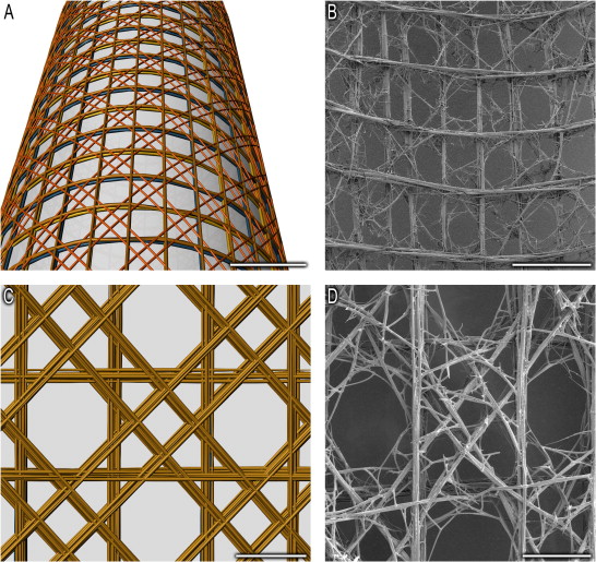
Fig. 7. The checkerboard-like structure of Euplectella aspergillum. A, B) Model of the pattern. B, D) Scanning electron microscopy image of a sample of Euplectella aspergillum. (Weaver et al., 2007).
Spicule Ctructure of Tethya aurantia
The sessile demospongiae sponge Tethya aurantia, commonly known as the orange puffball sponge, exhibits great buckling resistance due to its spicules design and layout (Fig. 8). The sessile creatures live where strong currents are continuously flowing; therefore, the spicules of the sponge have formed to provide the sponge with greater stiffness. The spicules of T. aurantia are axisymmetric and tapered along their length and approximately 35 μm thick, and 2 mm long. The non-laminated silica rods are called strongyloxea (Sxa) (Fig. 9). Additionally, the tapered ends are uniform between different Sxa’s. The number of spicules increases with the stiffness of the sponge. The sponge can maintain its shape even though it is subjected to forces from waves and currents in the coastal environment. The Sxa are staggered in bundles that radially stretch out from the center of the sponge to the surface (Fig. 10A). A cross-section of each bundle contains about 50 Sxa’s. The Sxa’s in each bundle are separated by a small amount of spongin (Fig. 10B and C). Spongin is much softer compared to Sxa’s (Monn & Kesari, 2017).

Fig. 8. Image of the orange puffball sponge (Monterey Bay Aquarium, 2024).

Fig. 9. Strongyloxea profile. Micrograph of several Sxa’s. [Adapted from Monn & Kesari, 2017].
When external force is applied to the sponge, it is transmitted by the spongin to the Sxa’s as tractions on the surface. These forces are then localized at the tapered ends of the Sxa’s (Fig. 10D and E). The mechanical stress is not distributed along the entire length of an Sxa. When axial stress is applied to the end of the bundles, a single spicule would act as a column and rotate. However, since the spicules are in a bundle, they prevent each other’s rotations leading to a static equilibrium (Fig. 10F). Therefore, using the Euler-Bernoulli beam theory, when the axial forces are applied to the end of an Sxa, the Sxa cannot move perpendicularly to its axis. This model is called the simply supported column (Fig. 10G). The transverse deflection, w, can be modeled by the differential equation:

Eq. 4. Transverse deflection of Sxa.
For all of z in (0, L), length as L of the spicule, and the boundary conditions:

Eq. 5. Boundary condition for the transverse deflection, w, of Sxa.

Eq. 6. Boundary condition for Youngs modulus, E, and the second moment of inertia, I, in Sxa.
Where P is the axial compression, E is Young’s modulus (stiffness of the material), and I the second moment of area. E is a constant and I(z) =πr(z)4/4. The r(z) is the cross-section radius of the Sxa. The buckling strength of an Sxa is the smallest axial compression (P) for a solution in Equations (4-6) that exists when w does not equal 0 and z is in [0, L]. When the Sxa profile is compared to other profiles such as semi-lapsed, triangular, and the Clausen profile (a model that is optimal for maximizing the resistance to buckling), the Sxa was shown to follow the Clausen profile, the best one (Fig. 11). Thus, the tapered ends of the Sxa’s provide for a 33% enhancement compared to a typical Clausen -profile cylinder in buckling strength. (Monn & Kesari, 2017).
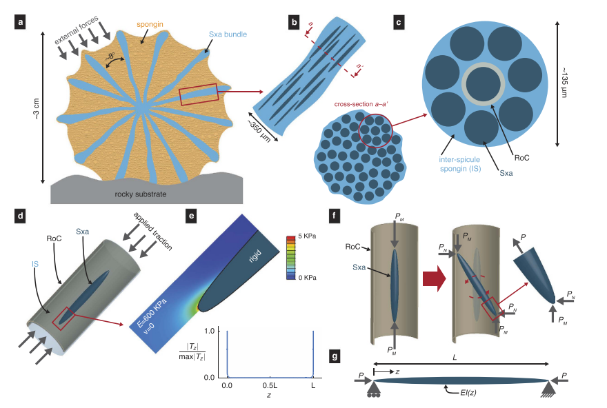
Fig. 10. Arrangement of Sxa’s in Tethya aurantia. A) Cross-section of the sponge showing the bundles of spicules radiating from center to outer edge. B, C) A bundle of Sxa’s in gray separated by the spongin in blue. D) model of an Sxa with applied traction to the ends. E) Spicule with stress applied showing how all the stress is distributed at the end of the spicule. F) Force applied to the end of the Sxa causes it to rotate but other spicules around stop the rotation. G) Model of simply supported column. (Monn & Kesari, 2017).

Fig. 11. An Sxa taper compared to other shapes. A) Four different column profiles. B) Best fit profiles for the different shapes. Sxa’s are shown as gray squares. (Monn & Kesari, 2017).
Holdfasts
Holdfasts are the root system that maintains the sea sponges in their sessile position. There are two predominant types of holdfasts. One is made up of many small anchor hooks attaching the body of the sponge to the base location, while the other is made of one strong anchor embedded in the seafloor (Fig. 12).
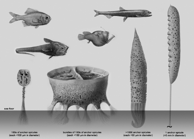
Fig. 12. Holdfast diversity in hexactinellid sponges. From left to right: Hyalonema, Chaunangium, Semperella, Monorhaphis. (Weaver et al., 2010a).
Holdfast of Euplectella aspergillium
Most Porifera use a system of hundreds to thousands of miniscule anchor spicules to attach. These are long, thin fibers which are about 3 times thinner than human hairs. Euplectella Aspergillum, a hexactinellid, has anchors roughly 10 cm long and 50 μm in diameter. Like most “above-ground” spicules, these anchor spicules are laminated and have 10-50 layers that decrease in thickness from core to periphery and are separated by organic interlayers. The layers are made of amorphous hydrated silica and surround a solid silica core. The holdfast spicules were also found to have recurved barbs (like thorns) coming from the spicules. While it is well known that laminated structures increase the amount of work required for fracturing in anchor spicules, Micheal Monn et al. built a model to analyze the strength of this specific root structure (Monn et al., 2015). In the model, the functionality of spicules was measured by load capacity (tensile force transmitted by surface barbs without failing). It was found, based on Bazant’s theory of stress redistribution and a derived equation for bending moment based on silica cylinders around the core, that load capacity always increases relative to an anchor spicule's thickness. In fact, thickness and number of silica cylinders are linearly related. The model finds that the sponges optimize their spicule design to increase load capacity, not even accounting for the progressive failure of laminated spicules, rather than a clean break (Monn et al., 2015). This means that the found load capacity is lower than the true support of anchor spicules. Analyses like these suggest that stronger materials could be built with composite beams, and sturdier foundations could be made mirroring the “barbed wire” roots of Porifera. Both would increase stress distribution.
Hexactinellid Sponge Monorhaphis chuni
Monorhaphis chuni is a species of sponge that has a giant anchoring spicule, known as the giant basal spicule (GBS). Unlike most sponges that anchor with multiple spicules, M. Chuni uses a single spicule.M. Chuni has a natural curve due to the exerted force from ocean currents. The curve creates a side of compression and a side of tension. For this reason, a cross-section of the sponge’s spicule reveals an asymmetric layer thickness surrounding the axial filament. The thickness of the layer is more on the compressive side and less on the tensile side. The tensile strength required to cause a fracture is proportional to h-1/2, where h is the thickness. Therefore, more tensile strength is required to fracture a thinner layer. Thus, the thinner layers are on the side experiencing more tension, providing the sponge with more strength to withstand the currents (Miserez et al., 2008). The thicker layers on the compressive side help to prevent buckling (Weaver et al., 2010b). Buckling occurs when a thin element, such as the large spicule, is exposed to a compressive axial force and begins to misshapen laterally once the critical magnitude of the force is reached (Monn & Kesari, 2017).
While the GBS is made of glass, it is not one rigid beam. When the micromechanical properties of the GBS were compared to that of a monolithic glass rod of the same dimensions, the anchor spicules had a 50% higher yield strength, a 45% increase in necessary stress for fracture (164 MPa for glass and 237 MPa for spicule), and exhibited a tenfold increase in toughness. The silica anchor spicule also had a less complete (or more fractured) breaking point, meaning that the force had been displaced over a greater surface area (Weaver et al., 2010a). In the aquatic environment, this could mean the difference between only suffering one deadly storm or surviving.
The interest in the giant basal spicule does not end there. The different concentric layers have different chemical makeups with different textures, they have zones of asymmetrical growth, and an external layer made of brown non-homogeneous organic content, while the inner layer is glassy (Pisera et al., 2021). Although not explored in depth, these different textures could be attempts by the sponge to create more distance between the layers and allow for more elasticity in the GBS.
Structural Biomimetics
Due to the long-term mechanical resilience exhibited by Porifera, like spicule strength and elasticity, they are an inspirational hub for the field of biomimetics. Already, there are widespread attempts at mimicking their lamination in structural materials, called spicule inspired structures (SIS). These are made of rigid resin (reinforced with SiO2) and “glued” with various organic mimics (E6000, superglue, etc.) (Sorour Sadeghzade, 2024).
The intrigue does not end there. The materials utilized by sponge structures including silica, spongin (collagen), calcium, and chitin, are known for their toughness and flexibility. Unconsciously replicating sponge mechanisms, some biomimetic labs are attempting to build a composite of collagen and chitin (Moon et al., 2019), which has been an important component in the holdfasts of sea sponges for millions of years (Ehrlich et al., 2013).
Fluid Dynamics in Sponges
Most sponges are suspension feeders (also filter-feeders), relying on both active pumping and passive ventilation to circulate flow within themselves. By directing streams through collars of microvilli, pseudopodia, and other filtration components, sponges can effectively extract bacteria, microorganisms, and other biological matter from the water, as well as take in oxygen for respiration. Interestingly, the sponges’ mechanism of extraction, specifically the collar-flagellated cells found in their aquiferous systems, exhibits a multitude of similarities with choanoflagellates, a group of aquatic unicellular colonial organisms. The shared structures, such as the flagellar vane (Fig. 13), support the superorganism evolutionary theory, where the Porifera phylum evolved from a colonial unicellular ancestor (Nielsen, 2019; Asadzadeh et al., 2020).

Fig. 13. Marked by white arrows is the flagellar vane of a choanoflagellate cell. These structures are also present on the flagella of sponge choanocytes (Mah et al., 2014).
Moreover, even sponges from distant phylogenetic lineages with different body plans have common cellular structures and organizations destined to direct filter-flow, reinforcing the common-ancestor theory (Lavrov et al., 2022). We will explore both the active and passive filtering methods in calcareous and hexactinellid sponges, highlighting their complex fluid dynamics.
Active Pumping
Suspension feeding sponges rely on a system of flagellated cells to direct water flow through the adequate filtering apparatus, which can vary widely from species to species. Ascon, leucon and sycon feeding chambers have varying morphology depending on evolutionary parameters: an evolutionary theory suggests that ascon-like filtration systems evolved into the three morphologies we know of today (Fig. 14). Nonetheless, a general aquiferous system and direction of fluid motion is shared across asconoid, leuconoid and syconoid forms (Fig. 14). In all three major categories of sponges, water enters ostia, the pores of sponges, through the single outlet (apopyle), reaching flagellated chambers. Here the water is filtered, as the flagella force it through collars and gaskets. Then, flow is directed towards the atrium, and then up and out through the osculum, the main opening at the top of the sponge (Asadzadeh et al., 2020).

Fig. 14. Diagrams of the sycon (S), ascon (A) and lycon (L) forms of sponge morphology (Asadzadeh et al., 2020). Blue arrows show the water path through the ostia into the chambers and out the apopyle. Notice the general direction of flow is the same despite the different feeding chamber morphologies.
About 10,000 choanocytes 3.5 µm in diameter were estimated to line an average-sized chamber in a syconoid sponge (Leys & Eerkes-Medrano, 2006). These cells pump water through hundreds of ostia, connecting outer water to the feeding chamber.
Choanocyte Cells
Choanocyte cells line the walls of the chambers of the sycon and lycon sponges, as well as the spongocoel –the central cavity– of the ascon sponge (Fig. 15). Their role is twofold: not only does the beating of the flagella serve as a pump to intake and expel water, but also favors retention of particles with backflow into the collar and strainer (Fig. 16).

Fig. 15. The structure of choanocyte cells surrounding the fenestrae in an asconoid sponge. Orange arrows indicate water flow. Ch – choanocyte, Co – collar, F – flagellum, Mv – microvilli, N – nucleus, St – strainer, V – flagellar vanes. [Adapted from Lavrov et al., 2022].
The filtration rate is significantly higher than the rate of flow entering the sponge through the ostium, underlining the importance of the re-filtering process (Fig. 16).

Fig. 16. The velocity field of a simulated flow within a chamber. Half of the ostium is on the right, marked by the dashed line. White arrows indicate the direction of water flow, suggesting backflow between each flagellum, as well as a larger backflow above the ostium, where the spacing between the flagella is large. [Adapted from Asadzadeh et al., 2020].
Pump Pressure Resilience
The characteristic curve is central to understanding how a sponge's pumping mechanism operates, particularly under different conditions of flow resistance. In a sponge without a gasket, the pumping unit would be each individual choanocyte. Each basic unit generates pressure and flow by working against the resistance in the system's canals. These pumping units work together in parallel, creating pressure that drives water through the collar filters (Asadzadeh et al., 2020). These pumps are characterized by two key factors:
Maximum Pumping Rate (Qmax): This is the flow rate when there is no pressure load.
Maximum Pressure (Pmax): The pressure generated at zero net flow.
The relationship between normalized pressure, represented by P = P / Pmax and normalized pumping rate, represented by Q = Q / Qmax , is linear, with the governing equation:

Eq. 7. Relationship between normalized pressure and pumping rate
The resistance of the system’s canals is modeled using Poiseuille flow, which applies to the movement of fluids through tubular structures. The pressure resistance in the system ( ) is defined as:

Eq. 8. Pressure resistance of the system assuming Poiseuille flow, where Rost = (128 /π)(μLost / Dost4), where Lost is the length of the ostium, Dost4 is its width, and µ is the dynamic water viscosity. Cpump is a characteristic constant of the pump, defined as Cpump = Pmax / Qmax (Asadzadeh et al., 2020).
When pressure is plotted against pump rate, the interaction of the pump’s characteristic curve (Eq. 7) and the system’s resistance curve (Eq. 8) defines the operating point of the sponge’s pumping unit. At this point, the optimal pumping rate, Q would be half of Qmax (Fig. 17).

Fig. 17. Pump and system characteristics for different pumping units and structures. The operating condition occurs at the intersection of these two curves, where the sponge operates most efficiently. (Asadzadeh et al., 2020).
Passive Ventilation
Many sponges have a particular skeletal geometry and structures to facilitate fluid flow through their fenestrae and guide it towards the osculum. This phenomenon is called passive ventilation and is employed by Hexactinellid sponges such as the Venus Flower Basket to facilitate filter feeding in the ocean depths, where currents are both rare and slow and choanocyte pumping is not as efficient. The advantages of the skeletal adaptations of the organism are twofold. They not only do reduce the drag experienced by the organism but also conveniently increase the water resistance time within the sponge, which allows it to extract particles more effectively (Falcucci et al., 2024).
Using computer fluid dynamics (CFD) simulation (Fig. 18), researchers isolated the passive ventilation of the sponge from its active pumping by modelling the structure without including motile choanocytes.

Fig. 18. A computer model of a Venus flower basket. (Falcucci et al., 2024).
To simulate a variety of flow conditions, a wide range of Reynolds numbers (Re), from 5 to 5000, were used during the research. These varying conditions simulate the different currents the sponge would encounter in its deep-sea environment, allowing a better understanding of the fluid dynamics around the structure at slow laminar flows and fast turbulent flows (Falcucci et al., 2024).
The Reynolds number is defined as:

Where u is the flow velocity, D is the outer diameter of the sponge, and v is the kinematic viscosity of the water.
The results of the simulation reveal that the skeletal structure of E. aspergillum naturally promotes an organized vertical flow of water through the body cavity, with fluid being drawn upward toward the osculum. This suggests that the sponge's design allows it to ventilate passively, without relying on active pumping mechanisms like flagella.
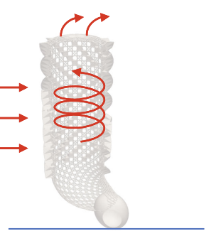
Fig. 19. A general diagram of how the skeletal structure of the sponge deviates currents (marked with red arrows). [Adapted from Falcucci et al., 2024].
The ridges of the sponge create a negative pressure gradient, effectively guiding water entering the body cavity towards the osculum. In Figure 20, we observe the statistical distribution of vertical component of flow velocity uz, depending on flow conditions with different Reynolds numbers.
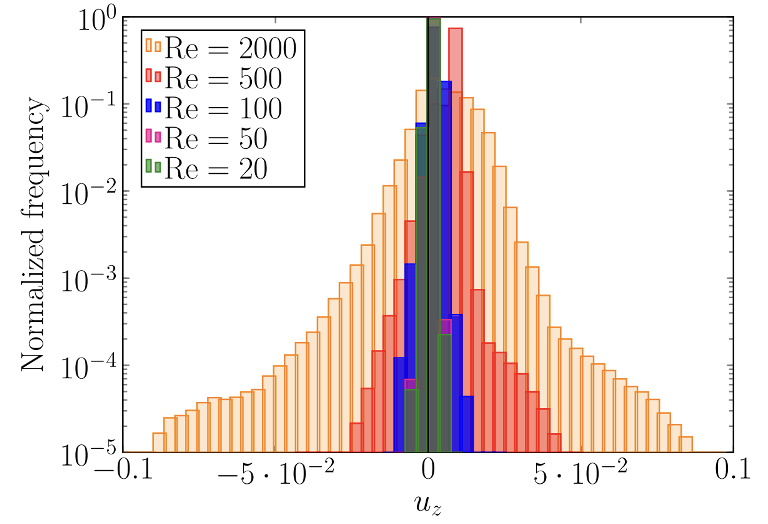
Fig. 20. Distribution of vertical component of flow velocity uz. The average values in lattice units for Re = 20, 50, 100, 500, and 2000 are 1.15×10⁻³, 0.68×10⁻³, 2.05×10⁻³, 6.64×10⁻³, and 5.82×10⁻³, respectively. (Falcucci et al., 2024).
With these results, the oscular flow rate Φ can be computed:

Eq. 9. Oscular flow rate, where A is the area of the cross-section of the osculum and <uz>osculum is the average flow exiting the osculum.
At low Reynolds numbers, which correspond to slow-moving ambient currents, the sponge’s passive ventilation system becomes more efficient. The study finds that at Re ~100, the sponge’s design is optimized for scavenging water and nutrients from its environment. At higher Reynolds numbers, the flow becomes more turbulent, but the sponge’s skeletal structure still manages to direct flow effectively toward the osculum, though less efficiently than at lower Re values (Falcucci et al., 2024).

Fig. 21. Percentage of flow exiting through the osculum at different Re numbers. [Adapted from Falcucci et al., 2024].
Optics
Light plays an important role in the function of numerous living organisms. From photosynthesis and vision to circadian rhythms, several biological processes rely on it. Sponges are no exception to the rule; they interact with light throughout their entire life cycle. This section exposes some of these curious interactions.
Led by Light
Each sponge begins its life as a larva. Unlike its parents, a parenchymellae larva is a tiny sphere of cells. The cells in its interior are embedded in a collagenous matrix, while the cells on its surface have cilia. Most cells have short cilia whose metachronal motion allows the larva to swim helicoidally. Two regions of the cell - the poles - have no cilia, however. The posterior one is particularly interesting because it is surrounded by a ring of pigments that is linked to longer cilia the larva uses to swim unidirectionally (Fig. 22) (Leys et al., 2002).

Fig. 22. Larvae of desmosponge Reneira imaged with light microscopy (A-C) and electron microscopy (D). A) A brood chamber (B ch) containing developing sponge larvae. Bar: 1 cm. B) Sponge larvae within the brood chamber. Notice the pigmented ring at their posterior pole (PRg). Bar: 1 mm. C) An individual larva with an anterior pole (AP) and a posterior pole (PP) with a pigmented ring and long posterior cilia (LPC). Bar: 100 µm. D) An individual larva fixed in its motion. Notice the short lateral cilia (SLC). (Leys & Degnan, 2001).
Since sponges spend their life attached to a substrate, larvae contribute to their dispersion as they are the only highly motile stage of the sponge life cycle. After being released from its parent, a larva needs to find a place suitable to metamorphose into an adult sponge, like under a rock or a reef flat (Leys & Degnan, 2001).
To accomplish this goal, the larva of the desmosponge Reneira that lives on the great coral reef begins its life by swimming upward towards the surface of the water. Geotaxis, its ability to use gravity to orient itself, is underpinned by a differential distribution of weight. The posterior end of the future sponge contains spicules that weigh it down. After its initial thrust into the seawater, the sponge embarks on its quest for settlement. Unfortunately, sponges have no neurons or gap junctions that could allow their cells to cooperatively decide where to make their new home (Leys & Degnan, 2001). The solution to this problem comes with light. Experiments on Amphimedon queenslandica, another demosponge, showed that the pigment ring surrounding its posterior pole contains two types of cryptochrome. Aq-Cry2, one of these molecules, absorbs light at 366-450 nm wavelengths, which coincides with one of the larva’s peaks of activity at 440 nm. Another peak at 600 nm suggests that this cryptochrome is only one of the elements responsible for the larval photokinetic response (Rivera et al., 2012). Regardless of the molecular details triggering the blue light response of sponge larvae, its mechanical aspects have been well studied. Leys demonstrated that exposure to light of appropriate wavelengths causes the long cilia on the posterior end of a sponge larva to strengthen and prepare for propulsion. It was proposed that the gradual exposition of the pigmented ring to the light source allows the larva to stir away from it. The exposition of one edge of the ring leads to ciliary motion that exposes the whole ring to the light source. As a result, the larva swims away from it (Fig. 23). This response persists in larvae up to their metamorphosis (Leys & Degnan, 2001).

Fig. 23. The gradual rotation of a sponge larva away from light. A) The exposure of the left side of the pigmented ring to light causes nearby cilia to beat. B) As a result, the larva turns its pigment ring towards the light source. C) The exposure of the ring causes all the cilia to stir the sponge away. (Leys & Degnan, 2001).
Made of Glass
After settlement and metamorphosis, the sponge becomes an adult whose skeleton is an arrangement of multiple silica spicules that have special optical properties. Kulchin et al. compared them to natural optical fibers. Using interferometry, the refractive index of 633 nm light in the 2-3 µm spicule core was measured to be 1.45-1.48. In contrast, the layer surrounding the core had a reflective index of 1.4. The layer around the latter has a refractive index increasing from 1.4 to 1.45 outwards. The indexes of the subsequent layers ranged from 1.4 to 1.35-1.39 and tended to decrease. While the initial refractive index corresponds closely to the one of fused silica, it decreases due to an increase in protein content. Similarly, the index of the second outer layer grows due to an increase in silica content, which makes it similar to an interface. The refractive index of the next layers varies due to variation in their composition and thickness. This structure confers sponge spicules optical fiber properties (Kulchin et al., 2009). Whenever light is incident upon an interface of two layers, if the refractive index of the outer layer is lower than that of the inner layer, there exists a critical angle. Exceeding this angle, an incident ray undergoes complete internal reflection and stays trapped in the spicule (Fig. 24).

Fig. 24. A multilayer optical waveguide model. Sponge spicules have a similar structure but with more layers with different refraction indexes. (Kulchin et al., 2009).
Nevertheless, some properties of spicules distinguish them from artificial waveguides. Spicules have a conical shape and contain a range of different materials and compounds (Kulchin et al., 2009). While the overall shape of a spicule may affect its properties and allow it to act as a lens (Voznesenskiy et al., 2010), for example, light is mainly modified by the ultrastructure of the spicule and its chemical characteristics. In other words, spicules are one-dimensional photonic crystals (Kulchin et al., 2009).
Observing the transmission spectrum of a spicule is a good starting point to understand the optical phenomena that happen in it. The transmission spectrum of a spicule from Hyalonema sieboldi, a hexactinellid, shows that the transmitted wavelengths are between 615 and 1310 nm (Fig. 25). Setting a lower and an upper limit to the transmitted wavelengths, the spicule acts as high-pass and low-pass filter, respectively. There are minima at 770, 960 and 1150 nm, which correspond to the absorption wavelengths of water. Small quantities of which are trapped in the spicules. Maximum transmission of 40-60% is observable around the 900 nm wavelength in the 1080-1100 nm range of maximum transmission. In fact, long spicules illuminated from one end display a gradient of color form white to red, which is explained by the transmission of longer wavelengths (Müller et al., 2006).

Fig. 25. Intensity of the incident light (magenta) and the light transmitted through a spicule of Hyalonema sieboldi (green) plotted against the wavelength. [Adapted from Müller et al., 2006].
This transmission spectrum depends on the absorption and scattering that happen within the spicule. Generally, these phenomena can be described in terms of the modes that can travel through an optical fiber. Each mode is a solution to the wave equation that is orthogonal to all other solutions. The latter means that it cannot interfere with other modes. Modes are simply perpendicular electromagnetic waves travelling through an optical fiber (Dändliker, 1999). When the optical parameters fall within the range typical to spicules only a small number of modes can exist within the core of the spicule. Several modes are leaky; they tend to escape.
Other optical effects happening within spicules are more exotic. Their structure creates photonic bandgaps (Kulchin et al, 2009). In brief, the structure causes destructive interference of certain wavelengths, which forbids their existence within the fibre (Ramaswami et al., 2010). This effect serves as the basis for two other properties. Spicules become able to conduct light in single and Bragg modes. The former means that light travels in a straight line within the fiber, and the latter that light undergoes diffraction. Lastly, spicules also act as free strand fibers, which are type of photonic crystals that redistribute the intensity of the incoming light over a wider range of wavelengths. The resulting distribution of light intensity is called a supercontinuum (Kulchin et al., 2009).
Despite their wonderous optical properties, spicules remain a biological conundrum. The concentration of silicium required for their optimal growth - between 5 and 100 µM - is significantly larger than its present concentration in the ocean - less than 3 µM (Müller et al., 2006). This discrepancy is due to the conservation of a silicium-fixation systems from an ancestor who lived in the pre-Tertiary when silicium was significantly more abundant (Maldonado et al., 2011). Thus, the conservation of silica spicules should provide some adaptive advantages superior to the potential benefits of their replacement (Müller et al., 2006). Some of these may be related to optics.
No Nerves, no Trouble
Two main hypotheses attempt to explain the use of the optical properties of spicules.
The first one addresses the lack of neurons in the sponge phylum. Since these animals have no nervous system but need to coordinate their rudimentary bodies, a mechanism should allow information to travel between their cells. Müller et al. suggested that light may be corner stone of their intercellular communication. His team identified a luciferase (an enzyme catalyzing bioluminescence) and a luciferin-regenerating enzyme (an enzyme regenerating the pool of the reactant used by the luciferase) in Suberites domuncula, a desmosponge. They hold that this pair of molecules forms an emitting system. If it were linked with the transmitting system formed by the network of spicules and a detecting system formed by a yet undiscovered pigments, the sponge could have a functional internal communication system. There is one caveat to this theory, some deep-sea sponges host bioluminescent symbiotic algae and bacteria. The light they produce may interfere with the cell-to-cell communication within the sponge (Müller et al., 2009). The problem worsens when one considers that the depths of the ocean are swarming with bioluminescent plankton that emits light upon collision with obstacles (Johnsen et al., 2012).
Voznesenskiy et al. proposed an alternative hypothesis that considers the interactions of deep-sea sponges with symbionts and plankton. The team found that the spicules of Pheronema raphanus, a desmosponge, function as short-focus lenses. Their arrangement within the organism concentrates light on the region of the sponge containing the highest concentration of organic matter, and their ends have traces of chlorophyll. Knowing that P. raphanus lives in symbiosis with cyanobacteria that cover between 10 and 30 percent of its energetic needs, the researchers proposed that these spicules may direct light on these photosynthetic symbionts. After observing another type of spicules at the surface of the sponge, the team concluded that they may stimulate bioluminescence from free plankton, which would also be collected (Voznesenskiy et al., 2010).
Optical Adaptations in Sponges
Sponges live in a multitude of habitats for which they developed a multitude of adaptations. Several of these adaptations, such as pigment-regulated cilia in larvae and silica spicules in adults, rely directly on light. Phenomena as varied as absorption, transmission, total internal reflection, photonic bandgaps, and single mode travelling regimes happen in sponges and help these animals to perform actions ranging from orientation to feeding.
Conclusions
In essence, the physical principles of the sponges’ environment have shaped their unique characteristics. These sea-dwelling creatures have evolved to form complex skeletal structures of spicules to withstand the forces of the ocean’s currents. One striking instance is the Euplectella aspergillum sponge evolving to have a hieratical spicule formation with maze-like ridges that allow water flow and prevents uproot of the sessile creature. Furthermore, each sponge has a root structure that anchors it to the seafloor. The structural components that form sponges have inspired scientists to create tough and flexible materials. The laminated spicule of sponges is the future of weather-resistant civil engineering due to the stress displacement and flexibility of the pattern of the material. On a different note, sponges have unique fluid dynamics capabilities that allow them to filter feed. Sponges that exhibit active ventilation have specialized cells to direct water through their filtering apparatus. Additionally, some sponges, such as the Venus flower basket, use their unique skeletal structure to direct the water flow within themselves. Finally, sponges utilize light to guide their movement through the water as larvae, and their spicules exhibit unique optical properties. Sponges can have spicules that display optical properties due to the varying refractive indexes of their multiple layers. Once the light enters the spicule it can remain trapped if the internal reflection exceeds a certain angle. Sponges feature many different characteristics from fluid flow to optics that allow them to thrive as sessile creatures.
References
References
Asadzadeh, S. S., Kiørboe, T., Larsen S. P., Leys, S., Yahel, G. Walther, J. (2020). Hydrodynamics of sponge pumps and evolution of the sponge body plan eLife 9:e61012
https://doi.org/10.7554/eLife.61012
Dändliker, R. (1999, 1999). The concept of modes in optics and photonics.Technical Digest Series Education and Training in Optics and Photonics,
Ehrlich, H., Kaluzhnaya, O. V., Tsurkan, M. V., Ereskovsky, A., Tabachnick, K. R., Ilan, M., Stelling, A., Galli, R., Petrova, O. V., Nekipelov, S. V., Sivkov, V. N., Vyalikh, D., Born, R., Behm, T., Ehrlich, A., Chernogor, L. I., Belikov, S., Janussen, D., Bazhenov, V. V., & Wörheide, G. (2013). First report on chitinous holdfast in sponges (Porifera). Proc Biol Sci, 280(1762), 20130339. https://doi.org/10.1098/rspb.2013.0339
El-Bawab, F. (2020). Chapter 4 - Phylum Porifera. Invertebrate Embryology and Reproduction, 106-169. https://doi.org/https://doi.org/10.1016/B978-0-12-814114-4.00004-7.
Falcucci, G., Amati, G., Bella, G., Facci, A. L., Krastev, V. K., Polverino, G., Succi, S., & Porfiri, M. (2024). Adapting to the Abyss: Passive Ventilation in the Deep-Sea Glass Sponge Euplectella aspergillum. Physical Review Letters, 132(20). https://doi.org/10.1103/physrevlett.132.208402
Fernandes, M. C., Aizenberg, J., Weaver, J. C., & Bertoldi, K. (2021). Mechanically robust lattices inspired by deep-sea glass sponges. Nature Materials, 20(2), 237-241. https://doi.org/10.1038/s41563-020-0798-1
Johnsen, S., Frank, T. M., Haddock, S. H., Widder, E. A., & Messing, C. G. (2012). Light and vision in the deep-sea benthos: I. Bioluminescence at 500-1000 m depth in the Bahamian islands. The Journal of Experimental Biology, 215(19), 3335-3343. https://doi.org/10.1242/jeb.072009
Kulchin, Y. N., Bezverbny, A. V., Bukin, O. A., Voznesensky, S. S., Galkina, A. N., A.L., D., & I.G., N. (2009). Optical and nonlinear optical properties of sea glass sponge spicules. In W. E. G. Müller & M. A. Grachev (Eds.), Optical and Nonlinear Optical Properties of Sea Glass Sponge Spicules (Vol. 47, pp. 315-340). Springer-Verlag. https://doi.org/10.1007/978-3-540-88552-8
Lavrov, A. I., Bolshakov, F. V., Tokina, D. B., & Ereskovsky, A. V. (2022). Fine details of the choanocyte filter apparatus in asconoid calcareous sponges (Porifera: Calcarea) revealed by ruthenium red fixation. Zoology, 150, 125984. https://doi.org/10.1016/j.zool.2021.125984
Leys, S. P., Cronin, T. W., Degnan, B. M., & Marshall, J. N. (2002). Spectral sensitivity in a sponge larva. Journal of Comparative Physiology A, 188(3), 199-202. https://doi.org/10.1007/s00359-002-0293-y
Leys, S. P., & Degnan, B. M. (2001). Cytological basis of photoresponsive behavior in a sponge larva. The Biological Bulletin, 201(3), 323-338. https://doi.org/10.2307/1543611
Leys, S.P., Matveev, E., Suarez, P.A. et al. Models of flow through sponges must consider the sponge tissue. Nature 603, E23–E25 (2022). https://doi.org/10.1038/s41586-021-04380-8
Leys, S. P., & Eerkes-Medrano, D. I. (2006). Feeding in a Calcareous Sponge: Particle Uptake by Pseudopodia. The Biological Bulletin, 211(2), 157–171. https://doi.org/10.2307/4134590
Maldonado, M., Navarro, L., Grasa, A., Gonzalez, A., & Vaquerizo, I. (2011). Silicon uptake by sponges: A twist to understanding nutrient cycling on continental margins. Scientific Reports, 1(30), 1-8. https://doi.org/10.1038/srep00030
Miserez, A., Weaver, J. C., Thurner, P. J., Aizenberg, J., Dauphin, Y., Fratzl, P., Morse, D. E., & Zok, F. W. (2008). Effects of Laminate Architecture on Fracture Resistance of Sponge Biosilica: Lessons from Nature. Advanced Functional Materials, 18(8), 1241-1248. https://doi.org/https://doi.org/10.1002/adfm.200701135
Monn, M. A., & Kesari, H. (2017). A new structure-property connection in the skeletal elements of the marine sponge Tethya aurantia that guards against buckling instability. Scientific Reports, 7(1), 39547. https://doi.org/10.1038/srep39547
Monn, M. A., Weaver, J. C., Zhang, T., Aizenberg, J., & Kesari, H. (2015). New functional insights into the internal architecture of the laminated anchor spicules of <i>Euplectella aspergillum</i>. Proceedings of the National Academy of Sciences, 112(16), 4976-4981. https://doi.org/doi:10.1073/pnas.1415502112
Moon, H., Choy, S., Park, Y., Jung, Y. M., Koo, J. M., & Hwang, D. S. (2019). Different Molecular Interaction between Collagen and α- or β-Chitin in Mechanically Improved Electrospun Composite. Mar Drugs, 17(6). https://doi.org/10.3390/md17060318
Monterey bay aquarium. (2024). “Orange Puffball Sponge.” Montereybayaquarium.Org, www.montereybayaquarium.org/animals/animals-a-to-z/orange-puffball-spon….
Müller, W. E., Kasueske, M., Wang, X., Schröder, H. C., Wang, Y., Pisignano, D., & Wiens, M. (2009). Luciferase a light source for the silica-based optical waveguides (spicules) in the demosponge Suberites domuncula. Cellular and Molecular Life Sciences, 66, 537-552. https://doi.org/10.1007/s00018-008-8492-5
Müller, W. E., Wendt, K., Geppert, C., Wiens, M., Reiber, A., & Schröder, H. C. (2006). Novel photoreception system in sponges? Unique transmission properties of the stalk spicules from the hexactinellid Hyalonemasieboldi. Biosensors and Bioelectronics, 21(7), 1149-1155. https://doi.org/10.1016/j.bios.2005.04.017
Nielsen, Claus. (2019) Early animal evolution: a morphologist's view. R. Soc. Open Sci. 6: 190638. https://doi.org/10.1098/rsos.190638
Palumbi, S. R. (1984). Tactics of acclimation: morphological changes of sponges in an unpredictable environment. Science, 225(4669), 1478-1480. https://doi.org/10.1126/science.225.4669.1478
Pisera, A., Łukowiak, M., Masse, S., Tabachnick, K., Fromont, J., Ehrlich, H., & Bertolino, M. (2021). Insights into the structure and morphogenesis of the giant basal spicule of the glass sponge Monorhaphis chuni. Frontiers in Zoology, 18(1), 58. https://doi.org/10.1186/s12983-021-00440-x
Ramaswami, R., Sivarajan, K. N., & Sasaki, G. H. (2010). Chapter 2 - Propagation of signals in optical fiber. In R. Ramaswami, K. N. Sivarajan, & G. H. Sasaki (Eds.), Optical Networks (Third Edition) (pp. 47-112). Morgan Kaufmann. https://doi.org/https://doi.org/10.1016/B978-0-12-374092-2.50010-2
Rivera, A. S., Ozturk, N., Fahey, B., Plachetzki, D. C., Degnan, B. M., Sancar, A., & Oakley, T. H. (2012). Blue-light-receptive cryptochrome is expressed in a sponge eye lacking neurons and opsin. The Journal of Experimental Biology, 215(8), 1278-1286. https://doi.org/10.1242/jeb.067140
Sorour Sadeghzade, N. F., Ajinkya Nene, Fariborz Tavangarian. (2024). Biomimetic 3D printed spicule-like composite of organic material/rigid resin cylinders with highly enhanced strength and toughness.
Uriz, M. J., Turon, X., Becerro, M. A., & Agell, G. (2003). Siliceous spicules and skeleton frameworks in sponges: origin, diversity, ultrastructural patterns, and biological functions. Microsc Res Tech, 62(4), 279-299. https://doi.org/10.1002/jemt.10395
Voznesenskiy, S. S., Kulchin, I., N., Galkina, A. N., & Sergeev, A. A. (2010). Morphological, optical, and structural characteristics of glass sponge spicules and the photoreceptor hypothesis of their survival. Biophysics, 55(1), 87-91. https://doi.org/https://doi.org/10.1134/S000635091001015X
Wang, X., Liu, A. G., Chen, Z., Wu, C., Liu, Y., Wan, B., Pang, K., Zhou, C., Yuan, X., & Xiao, S. (2024). A late-Ediacaran crown-group sponge animal. Nature, 630(8018), 905-911. https://doi.org/10.1038/s41586-024-07520-y
Weaver, J. C., Aizenberg, J., Fantner, G. E., Kisailus, D., Woesz, A., Allen, P., Fields, K., Porter, M. J., Zok, F. W., Hansma, P. K., Fratzl, P., & Morse, D. E. (2007). Hierarchical assembly of the siliceous skeletal lattice of the hexactinellid sponge Euplectella aspergillum. Journal of structural biology, 158 1, 93-106.
Weaver, J. C., Milliron, G. W., Allen, P., Miserez, A., Rawal, A., Garay, J., Thurner, P. J., Seto, J., Mayzel, B., Friesen, L. J., Chmelka, B. F., Fratzl, P., Aizenberg, J., Dauphin, Y., Kisailus, D., & Morse, D. E. (2010a). Unifying design strategies in demosponge and hexactinellid skeletal systems. Journal of Adhesion, 86(1), 72-95. https://doi.org/10.1080/00218460903417917
Mah, J. L., Christensen‐Dalsgaard, K. K., & Leys, S. P. (2014). Choanoflagellate and Choanocyte Collar‐flagellar systems and the assumption of homology. Evolution & Development, 16(1), 25–37. https://doi.org/10.1111/ede.12060