
Table of Contents
On this page
- Abstract
- Introduction
- The Auditory System
- The Somatosensory System
- Thermoregulation
- Cancer-resistant Cell Mechanics
-
The Cardiac Physiology and Biomechanical Adaptations
- Cardiac Size, Function, and Biomechanics
- Stress Response and Contractile Mechanics
- Ventricular Stiffness and Material Properties
- Biomechanical Testing and Stress-Strain Relationships
- Ventricular Mechanical Properties
- Histological Composition of the Naked Mole-Rat Cardiomyocytes
- Evolutionary Adaptation
- Conclusion
- References
Keywords: naked mole rat, subterranean environment, extended phenotype, energy efficiency, cardiovascular biomechanics, thermoregulation, evolutionary adaptation, sensory systems, hypoxia tolerance
Abstract
The naked mole rat is an ‘anomaly’ among mammalian species. It has adapted so well to its subterranean environment that it has extended key biological functions to its surroundings. Thus, its environment has become an extension of its phenotype. Deprived of light sources, this species has developed highly specialized auditory and somatosensory systems that employ its tunnel system to amplify sound and tactile stimuli. Their underground environment has forced the development of a unique thermoregulatory system that depends on the insulation of the tunnels to maintain optimal body temperature in the absence of subcutaneous fat. The mechanical properties of its cancer-resistance demonstrate the ways in which its habitat has shaped its cellular resistance against damage and stress. Finally, the hypoxic characteristic of its tunnel system has allowed its heart to function with reduced consumption of energy and oxygen. Ultimately, this paper explores the ways in which the naked has intertwined its physiology and anatomy with its environment, effectively extending its phenotype to the physical world.
Introduction
The naked mole rat (Heterocephalus glaber) belongs to the Ctenohystrica supraorder of rodents. This supraorder contains the two superfamilies Phiomorpha and Caviomorpha. The Phiomorpha are native to central and southwestern Africa and consists of species such as the naked mole rat, African mole rat, and porcupines (Lewis et al., 2016) (Holmberg, 2022). Although the naked mole rat was originally classified within the family Batheyergidae with the African mole rat species, it now resides in its own family Heterocephalidae (Figure 1). The change is the result of research done by Faulkes and his colleagues in 2004 (Faulkes et al., 2004). Their research discovered that the naked mole rat diverged 31 million years ago, before the African mole rat species. While they may be different families, both Batheyergudae and Heterocephalidaes are contain eusocial species (Lewis et al., 2016).

Fig. 1. The table depicts the phylogenetic relationship of the different rodent species. All numbers depict the millions of years ago when the divergence occurred (Lewis et al., 2016).
The naked mole rat possesses brown-pink loose skin and sparse tactile hairs (Buffenstein et al., 2011). They have powerful incisors dedicated to digging, which can be closed behind their lips to prevent soil from entering their mouths. Moreover, the naked mole rat thrives in complex hypoxic burrow tunnel systems. Each burrow contains specialized chambers for nesting, food storage, and toileting, and workers excavate soil in an assembly-line style, passing dirt back through the tunnels (Figure 2) (Naked Mole Rat - Zoo Atlanta, n.d.).

Fig. 2. The complex tunnel system containing specialized burrows that the naked mole rat builds and resides within (Parsons, 2012).
Naked mole rats form colonies which typically average around 70 members but can reach up to 300 members. These colonies include a single queen and various workers and breeders. The queen is the only breeding female in the colony. Her reproductive cycle is approximately 70 days, and she can produce a new litter every 80 days, resulting in up to five litters per year. In contrast, other colony members help care for the young, maintain the burrow system, and search for food, ensuring the stability and success of the colony (Naked Mole Rat - Zoo Atlanta, n.d.).
The naked mole rat feeds on nutritious roots and tubers found in arid regions and occasionally practices coprophagy, feeding on feces, to maintain gut microbial balance. In short, their diet includes various fruits, vegetables, and specially formulated mixtures to meet their nutritional needs (Naked Mole Rat - Zoo Atlanta, n.d.).
The Auditory System
The subterranean niche occupied by the naked mole rat is nearly devoid of light sources, which forces this animal to rely on its other physical senses, notably its auditory system. The naked mole rat has not only structurally modified its ears to suit its needs: it has also integrated its tunnel system to better its use and detection of sound.
The Structure of the Ear
While the structure of the naked mole rat’s ear is minimalistic, it is perfectly suited to its subterranean, low-noise environment (Mason et al. 2016). The external portion of the ear lacks pinnae, or external ear flaps (Mason et al. 2016), which has the function of funneling and amplifying sound waves into the ear canal (Jahchan et al., 2020). Instead, the naked mole rat possesses a small bump at the opening of the canal (Mason et al. 2016), clogged by hairs and earwax as seen in Figure 3 below. The blockage dampens incoming sound waves. Mammals without pinnae need a system to achieve collection and amplification of sound waves. The tunnels where naked mole rats reside fulfills this role and compensate the lack of a biological structure. They act like waveguides by amplifying acoustic waves traveling through their barriers (Barker et al. 2019).

Fig. 3. The figure shows the lack of pinnae, or sound capturing structures of the naked mole rat. It further illustrates the way in which the opening to the ear canal is clogged with hairs and earwax, a feature that serves to protect the ear from debris but ultimately dampens incoming sound waves (Barker et al. 2019).
The middle ear structures are condensed. The auditory bulla is compacted, and the malleus and incus bones are fused together (Mason et al. 2016). Thus, the middle ear’s compressed structure, which is responsible for the amplification and subsequent transmission of sound to the inner ear (Jahchan et al., 2020), reduces the naked mole rat’s sensitivity to sound (Mason et al. 2016). This anatomical compactness can be attributed to its inherent reduction of energy consumption, as the role of sound amplification is mostly unnecessary in the context of their acoustically conducive environment.
Furthermore, the inner portion of the ear lacks the intricacies that would allow an animal to have a large range of hearing (Mason et al. 2016). The cochlea, which is responsible for converting sound waves to nerve signals, has a very short contour length. The length of the cochlea correlates to the range of frequencies that can be heard by an animal (Jahchan et al., 2020). However, this animal does not need either a high sensitivity or a broad range, as it is only required to hear a specific, narrow range of frequencies. The dependence of the naked mole rat’s ear structure on its tunnel system exemplifies the ways in which its habitat has become an extension of its phenotype.
Range of Frequencies that Can be Heard by the Naked Mole Rat
As briefly mentioned above, a narrow range of frequencies can be detected by this animal. The naked mole rat can detect sounds between the frequency ranges of 125 Hz to 8 kHz, with their most sensitive frequency being found around 4 kHz (Barker et al. 2019). In addition to this narrow range of frequencies, a higher auditory threshold can be observed as well (Barker et al. 2019). This narrow frequency range and increased auditory threshold are unusual compared to other rodent species, as shown in Figure 4, which compares the hearing ranges of naked mole rats and other rodents (Barker et al., 2019).

Fig. 4. The auditory range versus frequency range of various rodents (Barker et al. 2019). This graph represents the naked mole rat through magenta squares. It shows that the naked mole rat has one of the narrowest frequency ranges compared to other rodents. Most species will exhibit peak sensitivity between 1 kHz to 10 kHz, whereas the naked mole rat is limited in its frequency range. It also has a significantly higher threshold, which indicates that it requires louder sounds to detect within its narrow range.
However, this small, low-frequency range has been documented in other subterranean species (Barker et al. 2019). The reason for this convergent adaptation lies in the physical properties of sound propagation, as well as the ecological environment itself. Sound waves can be described as alternating compressions and rarefactions of particles travelling through a medium (Serway et al, 2019). The ability of a sound wave to propagate is based on its wavelength and the proposed medium. Since frequency and wavelength are inversely proportional, sound waves with lower frequencies will have longer wavelengths, which will experience less absorption when moving through dense mediums, like soil.

In equation 1, V represents the speed of sound, which is constant in a specific medium. λ represents the wavelength of the sound waves, or the distance between two successive wave peaks. ƒ represents the frequency of the sound, which is the number of peaks that pass a certain point in 1 second. ƒ is also equivalent to 1/T, where T is the time that is required for a sound wave to travel from peak to peak (Serway et al, 2019).
In the case of the naked mole rat, lower-frequency sound waves will propagate further distances than higher-frequency sound waves in its tunnels as they will be less attenuated or weakened by the surrounding environment (Barker et al. 2019). In other words, the energy in these sound waves will not be dampened by the surrounding soil. Conversely, higher frequency sound waves will do the complete opposite, as they will be easily dampened by the surrounding soil. The efficient propagation of lower frequency sounds allows the naked mole rat to communicate over long distances (Barker et al. 2019). Thus, the naked mole rat uses the physical properties of its environment to extend its hearing range.
The Vocalizations of the Naked Mole Rat
The naked mole rat is a highly vocal species that employs 25 distinct vocalization patterns in various social contexts. As expected, the frequency ranges of these patterns are suited to the propagation through their tunnel system, as seen in Figure 5 below.

Fig. 5. The histograms in the figure above plot the frequency of a sound as a function of the time required to make the sound. This figure shows that all the vocalization patterns lie within the narrow range of frequencies that are best heard by the naked mole rat.
Their complex eusocial structure, which includes a dominant queen that controls the breeding system, requires this large repertoire of vocalizations to communicate the needs of the colony (Barker et al. 2019).
For example, the soft chirp, the most common vocalization, plays a critical role in the identification and recognition of colony members. The ability to recognize members of their own colony is critical in a habitat devoid of light.
Other vocalizations are more context-specific, such as the toilet call. This vocalization is employed when a breeding individual is urinating and may represent a display of rank in the colony. This display of rank is not comparable to the boastful nature of humans. Instead, it is essential in maintaining order within the colony.
Courtship rituals also require unique vocal patterns. During male courtship songs, males will employ the mordent call to capture the attention of the queen. In the following phase, during mating, the queen and her mate will perform a vocal duet known as the V-trill. This type of vocalization plays a crucial role in facilitating reproduction and ensuring the continuation of the species.
Aggressive interactions also require their own set of vocalizations. These intense patterns include the grunt and loud chirp, both of which are employed during fights. However, if these aggressive encounters go too far, the scream is employed to express pain or injury.
The naked mole rat will even employ certain sounds to help navigate its tunnel system. For example, the tap is a sound that is made as the naked mole rat taps one of its paws on the tunnel floor to navigate the tunnels (Barker et al. 2019).
Finally, while dominant individuals do not necessarily have their own set of vocal patterns, they exploit the properties of sound propagation through tunnels to affirm their status in the established social hierarchy. Specifically, larger mole rats typically dominate their smaller counterparts. Because of their larger stature, they possess larger vocal apparatuses, which produce lower frequency sounds. Thus, researchers suggest that these lower-frequency sounds are more likely to elicit a response from other colony members, as their vocalizations travel more effectively through the tunnels. This relationship between size and response percentage is shown in the graph below (Figure 6) (Yosida et al. 2009).

Fig. 6. The graph shows the response percentage of naked mole rat members as a function of the weight of the member producing the vocalization. In the experiment conducted, naked mole rats of different weights were stimulated to vocalize. Then, the response of other members of the colonies were recorded as a percentage (Responsive naked mole rats/Total naked mole rats). It illustrates that larger mole rats will elicit higher levels of responses than smaller mole rats, as their low-frequency vocalizations will propagate better through the tunnel system (Yosida et al. 2009).
The chosen vocal patterns not only show the naked mole rat's dependence on vocal communication but also illustrate this species' use of its surrounding environment to extend its auditory capabilities and enhance essential social interactions. Their environment even extends the vocal abilities of its dominant members.
The Somatosensory System
The somatosensory system of the naked mole rat also manifests co-option of its environment. Its environment’s lack of abundant light has led to the tunnels of the naked mole becoming an extension of its somatosensory system. It has evolved specialized body hair and vibrissae that are able to detect intricate details about the environment that it interacts with. In turn, the tunnels are able to relay critical information through these vectors such as the orientation and position of the naked mole rat.
Body Hair
Contrary to its name, the naked mole rat is not entirely naked and has about 40 tactile hairs spread throughout its body (Crish et al., 2003). Hair does not adequately describe these structures as it more closely resembles vibrissae found on the snout of the naked mole rat. To navigate the long and complex networks of tunnels that the naked mole rat lives in, it requires an ability to sense its position and orientation. One method it uses to sense its direction is its usage of an increased sensitivity in its body hairs. These hairs are arranged in a grid-like pattern and when any mechanical force is applied to them, the surrounding nerves become stimulated (Crish et al., 2003). In conjunction with its tunnel, the increased sensitivity creates an extended phenotype that can detect both position and orientation. When individual hairs have been stimulated on naked mole rats, they have been shown to correctly orientate their snout to the direction of the stimulation at a rate of 95% (Figure 7) (Crish et al., 2003). This method of orientation allows the naked mole rat to perceive the direction it is walking in as well as surrounding objects in its tunnel.

Fig. 7. Recreation of video tapes of three different trials A, B, C where the naked mole rat had individual body hairs stimulated. The only difference in these trials was the rostro-caudal locus of the stimulated hair. The shadow shows the position of the naked mole rat prior to stimulation and the outline shows their position post stimulation. The small arrowhead in the top images shows the approximate rostro-caudal location of the stimulated hair. The arrow below shows the final turn angle the naked mole rat did in response to the stimulation (Crish et al., 2003).
Vibrissae
As a result of its thickness, the body hair of the naked mole rat resembles its vibrissae (Figure 8). However, it presents a slightly different relationship with the tunnel. Typically, vibrissae found on the snouts of animals are used by the somatosensory system to more precisely detect the texture of objects around it [hyperlink to whiskers and antennae, Hyperbook, 2022]. The naked mole rat’s diet consists of foraging for underground roots (nationalzoo.si. ed, 2024). Therefore, as it digs, it needs to be able to distinguish textures to discern food from soil. Via a more intricate nerve network, its vibrissae can not only detect general forces being applied but can even distinguish textures between objects (Park et al., 2003). The vibrissae-tunnel phenotype helps the naked mole rat distinguish food from soil because the tunnel has different textures which the vibrissae can detect.

Fig. 8. Scheme A shows the widely spaced body hair of the naked mole rat and B shows its vibrissae (whiskers) (Catania et. Al, 2002).
The Structure of Body Hair and Vibrissae
The structure of the naked mole rat’s body hair is more comparable to that of vibrissae than body hair found on other rodents. The similarity in structure allows for them to both have a relationship with their tunnel. One reason that their body hair can act in a similar way is that they are so sparsely spaced and are much thicker than body hairs of comparable rodents. Naked mole rat body hairs are on average 39 μm in diameter and their vibrissae are on average 38 μm diameter (see Figure 9) (Crish et al., 2003). This greater thickness leads to a higher level of stiffness akin to vibrissae. Higher stiffness causes a higher force being applied to the surrounding mechanoreceptors around the hair follicle, increasing sensitivity (Quist & Hartmann, 2012). The increased sensitivity of forces being applied to the body hair of the naked mole rat is how the body hair of the naked mole rat is so vibrissae-like.

Fig. 9. The ratio of the thickness of body hair and vibrissae of naked mole rats was found to be different to the ratio of rats. Instead, it was found to have the same thickness as their vibrissae (Crish et. Al, 2002).
Force Mechanics of Body Hair/Vibrissae
Although little research has been done on the exact force mechanics of the body hair of naked mole rats, since they are comparable in structure to vibrissae, it can be expected that they have similar mechanics. They do this by acting as a lever. When the hair/vibrissae bends, they generate forces and bending moments at their base (Quist & Hartmann, 2012). This greatly increases the sensitivity of the region in which the hair/vibrissae are present. These forces have been modeled and the external forces applied on the hair/vibrissae can be used to determine the forces which are transferred to the surrounding, nerve-containing tissue. The models assume a force normal to the hair/vibrissae, allowing for friction forces to be ignored, that can be broken into axial force (Fx), transverse force (Fy), and moment (Mz) which are all related to the angle from which the vibrissae are at rest (θp) (see Figure 10) (Quist & Hartmann, 2012). The forces and moments at the base of the vibrissae follicle, as a result of the impact of the vibrissae with an object, cause reactions in the surrounding nerve endings (Quist & Hartmann, 2012).

Fig. 10. Scheme B depicts how the forces (F) on the end of the vibrissae and body hair by an object are translated into forces at their base (Fx, Fy, Mz) which can later be converted into nerve signals. Scheme A depicts the amount of malformation from the interaction of the vibrissae with the object which is directly caused by the force vectors (Quist and Hartmann, 2012).
The Nerve Structure of Body Hair and Vibrissae
The structure of the vibrissae and body hair of the naked mole rat is not the only comparable phenotype it possesses. The naked mole rat’s brain has evolved to perceive the signals sent from both vibrissae and body hair. The superior colliculus is a section of brain that most mammals use for visual orientation (Crish, 2006). However, naked mole rats have evolved to use their superior colliculus for tactile orientation. When stimulated, both their vibrissae and body hair have been shown to send their signals to the superior colliculus where the signals are interpreted (Crish, 2006). However, the body hair does not contain all the types of nerves that the vibrissae does (Park et al., 2003). While greater thickness increases sensitivity to stimuli, it does not allow the body hair to detect properties that only the nerves in vibrissae can (Lewin, 2021). The body hairs of naked mole rats do not have circumferential nerve endings within their proximal dermal region whereas their vibrissae do (Figures 11 and 12) (Park et al., 2003). These nerve endings around the vibrissae are what allow the vibrissae to detect texture. They do this by using the changes in the applied forces as the vibrissae rubs against the bumps on the textured surface. They are sufficiently accurate with their vibrissae to detect shallow grooves spaced at 90 μm (Carvell, 1990). This level of accuracy means that the naked mole rat can distinguish 225 grit sandpaper which also has grains of 90 μm (Graham, 2024). The ability to detect texture as a result of different types of proximal nerve endings is the defining difference between the naked mole rats body hair and vibrissae.

Fig. 11. Scheme depicting the different structure of innervation present in the body hairs of the common mole rat (CMR) and rat compared to the innervation of the naked mole rat. Naked mole rats body hair contains a different innervation structure to other rodents' body hairs allowing them to more accurately detect stimulation (Park et al, 2003).

Fig. 12. Scheme depicting the innervation of the vibrissae of the naked mole rats compared to the common mole rat (CMR) and rat. The innervation of the vibrissae of the naked mole rat is very similar to that of other rodents and contain nerve endings which the body hairs do not (See figure 7 to compare) (Park et al, 2003).
The Function of the Naked Mole Rat’s Incisors
The naked mole rat’s body hair and vibrissae are not the only body parts that play a role in somatosensation. There is a large portion of their somatosensory cortex dedicated to the incisors. As shown in Figure 13, 1/3 of the somatosensory cortex is devoted to the incisors, along with a 1/4 devoted to the jaw muscles used for digging the soil (Buffenstein et al., 2021).

Fig. 13. Percentage of somatosensory cortex distributed to body parts of the naked mole rat (Buffenstein et al., 2021).
In addition, the naked mole rat has independently-moving incisors. The flexible mandibular symphysis (area along the midline of the lower jaw where the left and right halves of the jaw bones meet) allows the incisors to move independently (Catania and Remple, 2002). Its incisors play important roles especially when the juveniles are approximately 21 days. They spar with each other by locking their teeth to each other and play tug of war using their incisors to determine the hierarchical positions. The determination of hierarchical position is important as higher ranked naked mole rat pass on top of the naked mole rat with lower rank in their narrow tunnels (Buffenstein et al., 2021). Thus, it can be concluded that their narrow tunnels have led to the development in sparring among juveniles for hierarchical rank.
Furthermore, the naked mole rat has ever-growing incisors, like other rodent species. To offset the frequent usage of their incisors for digging, researchers suggest that the naked mole rats form digging chains, where multiple naked mole rats form a line to excavate the soil (see Figure 1). Thus, the independently-moving incisors and their constant eruption rate demonstrate the influence of their environment on their physical and behavioral traits to function in harmony.
Thermoregulation
The naked mole rat lives in a subterranean environment with constant temperature and limited food resources (Buffenstein et al., 2001). This environment led to one of its unique physiologies: its mechanism of thermoregulation (Buffenstein et al., 2001). While most mammals are endotherms, naked mole rats are poikilotherms (Figure 14) (Yahav & Buffenstein, 1991). The naked mole rat has poor insulative skin due to the limited amount of subcutaneous fat. Due to the naked mole rat’s small body size, its high surface area to volume ratio facilitates heat loss. Thus, utilizing the tunnel as its extended phenotype, the produced body heat is not retained but transferred to the environment (Daly et al., 1997). The naked mole rat’s unique thermoregulation mechanism is a result of adaptation to its subterranean environment.

Fig. 14. Description of 4 different types of thermoregulation. Poikilotherm is a thermal state where the animal has large range of body temperature and is heavily affected by the ambient temperature (Buffenstein et al., 2021).
The unique thermoregulation mechanism of the naked mole rat involves processes of heat and moisture exchange with their environment. The heat exchange between the naked mole rat’s metabolic heat and the tunnel occurs through conduction, convection, and radiation (Figure 15). For example, if the body temperature of a naked mole rat is higher than the environment, the body heat is transferred to the environment, and vice versa. Because the naked mole rat exchanges heat with the environment, its body temperature corresponds to the ambient temperature. As shown in Figure 16, the naked mole rat’s body temperature has a direct linear relationship to the ambient temperature (Buffenstein et al., 2021). The linear relationship implies that as the ambient temperature changes, their body temperature changes accordingly.

Fig. 15. The naked mole rat is poikilotherm, an organism which its body temperature is determined by the combined effects of metabolic heat production, heat loss through evaporative cooling, and heat exchange with the environment via thermal radiation, convection, and conduction (Buffenstein et al., 2021).
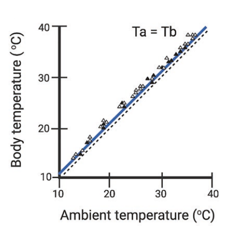
Fig. 16. Graph shows the effect of ambient temperature (Ta) on body temperature (Tb) of the naked mole rat (Buffenstein et al., 2021).
The body temperature of the naked mole rat is not only affected by heat exchange, but also the water exchange with its environment via evaporative cooling (Figure 15). The exchange of water between the naked mole rat and its environment depends on the vapor pressure gradient. The quantitative value of how easily the naked mole rat loses heat and water is expressed by wet thermal conductance. Due to its lack of subcutaneous fat and high surface area to volume ratio, the naked mole rat has 2-5 times higher water thermal conductance than similarly sized rodents, resulting in high heat and water exchange (Buffenstein et al., 2021).
The naked mole rat’s easily changing body temperature evolved them to withstand a large range of body temperatures. The naked mole rats can endure temperatures as high as 42°C, and as low as 12°C. Even after exposure to low temperatures for several hours, they can recover the optimal metabolism when they are exposed to a standard room temperature of 20°C-25°C. Even more surprisingly, they had no change in DNA fragmentation or increased rate of apoptosis even after long exposure to high temperatures. Despite their ability to withstand extreme temperatures, they have several strategies to maintain the optimal body temperature. They either seek heat from the environment or huddle among their colony by resting together in nests (Buffenstein et al., 2021).
To adapt to their environment with limited food, it is hypothesized that the naked mole rat has evolved to reduce the energy consumption for thermoregulation. Since the constant ambient temperature resembles its body temperature, evolving into a poikilotherm and utilizing the tunnel would be advantageous in terms of energy efficiency.
Cancer-resistant Cell Mechanics
The naked mole rat is known for its cancer resistance and longevity- up to 30 years of lifespan, which is about 9 times longer than the laboratory mouse. Due to such an advantageous trait, there has been ongoing research to identify the biological mechanisms that cause cancer-resistance in naked mole rats. However, the evolutionary adaptation that led to cancer-resistance in naked mole rats remains unknown (Makarova et al., 2022).
The naked mole rat’s subterranean environment, which protects it from predators, pathogens, and climatic extremes, was thought to be a contributing factor for its longevity and cancer resistance. However, according to research done by Kim (2011), the naked mole rat is shown to be not only resistant to spontaneous cancer, but also experimentally induced tumorigenesis. The naked mole rat’s resistance from induced tumorigenesis implies that a biological factor, rather than an environmental factor, could be the primary reason for its cancer resistance. In addition, according to Makarova (2022), upon oncogene and antigen treatment, mouse fibroblast cells transformed into malignant cells, while naked mole rat fibroblast cells showed no transformations. The result suggests that there is a biological factor contributing to cancer-resistance in the naked mole rat that is absent in mouse.
To further investigate, the naked mole rat fibroblast cells were experimented via atomic force microscopy (AFM), shown in Figure 17.

Fig. 17. Illustration of atomic force microscopy with variables used to calculate the rigidity of the cell. Atomic force microscopy is a scientific instrument that measures the rigidity of cells, hence providing information about the cell’s physical properties. Atomic force microscopy can collect data regarding the deformation of cells by obtaining the force curves of the cell surface cell topography (Makarova et al., 2022).
Figure 17 illustrates how the degree of cell deformation is quantified. From Equation 2, h is the distance between the cell and the atomic force microscopy probe. h=0 indicates a point where the cell is fully pushed by the probe. Effective Young’s modulus (E) is a variable that indicates cell stiffness. Z is the cell's vertical position when the force is applied by the atomic force microscopy tip to the cell. d is the amount of deflection of the atomic force microscopy tip, which indicates the force exerted on the cell. The bigger the Z and d, the greater force is applied to the cell. k is the spring constant of atomic force microscopy tip. Rprobe (Rcell)is the radius of the probe (cell) (Makarova et al., 2022).

Eq. 2. Equation is used to quantify the degree of cell deformation, or the stiffness of a cell when a certain amount of force is applied. Atomic force microscopy would provide values to calculate E, a variable that quantifies cell deformation (Makarova et al., 2022).
According to Makarova (2022), the atomic force microscopy results show that there was no statistically significant change in the deformation of cells of the naked mole rat fibroblast cells. On the other hand, there was statistically significant change in the cell mechanics of mouse fibroblast cells. The result implies that upon force, the naked mole rat fibroblast cells are more resilient to cell deformation compared to the mouse fibroblast cells. Thus, the fibroblast cells of naked mole rats are stiffer than those of mice. It is commonly accepted that softer cells are associated with cancer progression. Since the fibroblast cells of the naked mole rat are stiffer than those of mouse, the result suggests that the naked mole rat is less associated with cancer progression. Utilizing atomic force microscopy, the scientists were able to uncover the changes in the cell mechanics of the naked mole rat fibroblast cells under applied force, as an indication of the naked mole rat’s resistance to cancer (Makarova et al., 2022).
Even though research was able to uncover the possible biomechanics of their cells that may have an association with it being cancer-resistant (Seluanov et al., 2009), the evolutionary adaptation that led to such a trait is unknown. However, the hypoxia and food scarcity in the subterranean environment could be a contributing factor to their cancer-resistant phenotype. Such an environment forced them to have a slow metabolic rate for energy conservation, which reduced the production of reactive oxygen species, highly reactive molecules that are associated with aging and cancer. With low reactive oxygen species production, they would be less likely to age and have cancer, resulting in longevity and cancer-resistant phenotype (Buffenstein et al., 2021). Thus, their subterranean environment forced them to produce less reactive oxygen species that gave rise to such advantageous phenotypes.
The Cardiac Physiology and Biomechanical Adaptations
The naked mole rat exhibits high tolerance to hypoxic (low oxygen) environments and energy efficient metabolic strategies. The physiological adaptations also extend to their heart and circulatory system.
Cardiac Size, Function, and Biomechanics
One of the naked mole rat’s primary adaptations to its surroundings is the balance between heart size and contractility. The significance of this adaptation comes from the regulation of cardiac function in both normal and diseased hearts, which is influenced by biomechanics (Voorhees & Han, 2015). Cardiac measurements were obtained through an echocardiogram, which is a test that uses sound waves to produce images of the heart (Echocardiogram, 2023). These measurements showed that the ratio naked mole rat’s heart size relative to its body size is unlike the ratio in other mammals, such as the C57BL/6J mouse (Grimes et al., 2013). This finding suggests an increase in volume of the heart to pump blood, which can be linked to the pressure-volume relationship in cardiac physiology. This relationship examines how the pressure and volume of the heart's left ventricle fluctuate with each beat. This relation can be depicted using an examination of left ventricular pressure and volume with time, as well as the pressure-volume loop (Human Physiology Students from University of Guelph, n.d.).
Additionally, key biomechanical parameters such as fractional shortening, ejection fraction, and stroke volume describe how the heart converts mechanical energy into pumping. Fractional shortening measures the change in the dimension of the left ventricle during contraction of the heart. When factional shortening was measured in the naked mole rat, it was slightly lower than the mouse, as shown in Figure 18 (Grimes et al., 2013). Low fractional shortening indicates that the heart of the naked mole-rat undergoes less contractile deformation with each heartbeat, which can be considered an energy saving measure.
The naked mole rat has exceptionally low baseline heart function, which indicates that the heart beats at a slower pace than that of other rodents; nonetheless, this minimal degree of function is supported long into its age. Its heart rate is remarkably low (250 bpm) compared to mice (700 bpm), and its fractional shortening is lower by 28% than that of other rodents (Grimes et al., 2017).

Fig. 18. Comparison of mice to naked mole rats at baseline and after dobutamine-induced cardiac stress, echocardiography reveals that mice show higher left ventricular (LV) contractility. Echocardiogram time scale is in seconds. (Grimes et al., 2013).
As per the study conducted by Grimes, the thicker left ventricular walls in the naked mole rat are used for structural support. It also enables the heart to pump strongly so they can maintain proper functioning at low consumption of energy. This is a result of biomechanical optimization, in which the heart of the naked mole rat has adapted to manage copious quantities of blood being pumped by the heart at a slower rate than other animals, which reduces damage to the heart and maintains constant blood flow (Grimes et al., 2013).
The characteristics of the heart's size, function, and biomechanics are ecophysiological responses to their subterranean environment. They may also have some significance in the slight deterioration of heart function that occurs with aging (Grimes et al., 2017).
Stress Response and Contractile Mechanics
The heart of the naked mole rat showed an inability to enhance its output when put under stress via an administration of dobutamine (Figure 18) (Dobutamine Injection, 2024). Dobutamine is a drug used to treat heart failure via strengthening the heart muscle. This drug often increases cardiac activity and fractional shortening by activating the sympathetic nervous system and simulating the physiological effects of exercise or stress. However, in the naked mole rat, the effect was less apparent in comparison to mice, and contractility barely improved. This result indicates that its heart functions with a limited contractile reserve, as an alternative to maintain long-term efficiency in hypoxic environments (Grimes et al., 2013).
Ventricular Stiffness and Material Properties
According to the research established by Grimes, lower collagen fiber concentration in the naked mole rat’s heart is the main reason for its reduced ventricular stiffness, in comparison to the mouses heart. This decrease in collagen concentration has significant effects on the heart's capacity to pump blood during hypoxic stress, since it makes the heart more flexible and enables it to take up blood flow without using too much energy during contraction (Figure 15) (Grimes et al., 2013). This flexibility in the heart is essential to the survival of the naked mole-rat because it lowers the passive energy needed to fill the ventricles during diastole, which is the period of the heart's contraction during which blood fills the chambers (Grimes et al., 2013).

Fig. 19. The mouse and the naked mole rat (NMR) exhibit analogous left ventricular interstitial deposition of collagen (Grimes et al., 2013).
Biomechanical Testing and Stress-Strain Relationships
According to Grimes’s research, scientists conducted biomechanical studies on naked mole rat cardiac tissue to determine the relationship between stress, which is the internal force generated by heart tissue in response to deformation, and strain, which refers to the extent of that deformation. These studies employed the Fung-type strain energy density (W) as shown in equation 3, is a model used to explain nonlinear elastic behavior of biological tissues (Grimes et al., 2013).

The equation above demonstrates the strain energy density function. Q is a term that combines how the tissue stretches in two directions (circumferentially and lengthwise) and c is a constant derived from experimental data (Grimes et al., 2013).
Experiments have been conducted to assess the mechanical properties of the naked mole rat heart tissue. They found that the tissue had more compliant mechanical qualities than the mouse, which means that it is stretched and deformed easier under mechanical stress and strain (Grimes et al., 2013). To further test this, the heart tissue was subjected to biaxial tensile testing, where tissue samples were stretched in longitudinal and circumferential directions to see how the tissue responded. The increased deformation allows their hearts to handle changes in blood pressure and volume and to withstand the mechanical requirements of hypoxic environments (Grimes et al., 2013).
Ventricular Mechanical Properties
The mechanical properties of the naked mole rat’s heart reveals adaptations that allow it to manage stress without resulting in structural damage. To understand this, researchers used the Law of LaPlace (Equation) (Burkhoff, 2003). This principle is important because wall stress indicates how much force the heart muscle must handle when it contracts and relaxes, and higher wall stress usually leads to more strain on the heart (Vindis-Jesic et al., 2002). However, the naked mole rat myocardial tissue shows greater flexibility and compliance, allowing the heart to handle these pressures effectively during diastole (heart relaxation) and systole (heart contraction) (Grimes et al., 2013).

The calculations of the stretch ratios under diastolic pressures using the Law of Laplace, shown above, and diastolic dimensions are done to compare the mechanical properties under physiological conditions, (Grimes et al., 2013).
Equation 4 estimates how much stress (σ) is on the heart wall based on the blood pressure inside the heart (LVP), the radius of curvature of the heart (R), and the thickness of the heart wall (h)
When comparing the size of the naked mole rat’s heart to mice, the stress did not differ much between both animals (Figure 20) (Grimes et al., 2013). This shows additional evidence that the myocardial tissue in the naked mole rat can handle mechanical loads, such as blood pressure and volume changes.

Fig. 20. When comparing the left ventricular mechanics of the mouse and the naked mole rat (NMR), there seemed to be no apparent difference among both species (Grimes et al., 2013).
Hence, the findings show that the naked mole rat depends more on the passive mechanical properties of the heart instead of relying on increasing heart rate to cope with physiological stress. Passive mechanical tissue properties are major determinants of myocardial contraction, relaxation, and shape cardiac function (Emig et al., 2021). This regulation of mechanical stress may be a critical evolutionary adaptation.
Histological Composition of the Naked Mole-Rat Cardiomyocytes
According to Grimes et al. (2013), additional distinctions were observed at the cellular level. For example, naked mole rat cardiomyocytes, which are the contractile cells of the cardiac muscle, have a significantly greater cross-sectional area than those of mice (216 ± 10 µm² vs. 178 ± 7 µm²). This increase in cross-sectional area could improve the heart’s capacity to generate force per contraction, complementing the naked mole-rat's low heart rate and high cardiac output (Fig. 17) (Grimes et al., 2013).

Fig. 21. The cross-sectional areas of cardiomyocytes were bigger in the naked mole rat left ventricular (Grimes et al., 2013).
In addition, the arrangement of cardiomyocytes in the naked mole rat differs from that of mice since the naked mole rat’s cells are more circumferentially aligned, resulting in better force transmission and less strain throughout the heart muscle during contraction. Having said that, naked mole rats and mice share a similar cardiomyocyte configuration at the epicardium, which is the outermost layer of the heart. Naked mole rats exhibit a lower rotation of the fiber angle, meaning that their hearts do not need to twist as much to generate the force necessary for contraction. This suggests a more stable and efficient contraction of the heart while reducing strain on the cardiac muscle (Nakatani, 2011). In fact, this alignment of cardiomyocytes and lower rotation may be crucial to the naked mole rat's capacity to maintain effective ventricular function.

Fig. 22. Reduced fiber rotation of cardiomyocytes in the naked mole-rat left ventricular (Grimes et al., 2013).
Evolutionary Adaptation
The combination of improved mechanical properties and distinct histological traits demonstrates the naked mole rat's evolutionary adaptations to their subterranean environment. Furthermore, the bigger cardiomyocytes, along with their specific alignment, indicate that the naked mole rat’s heart is physically and functionally able to handle the difficulties of its environment (Grimes et al., 2013). By optimizing the mechanics of its heart and reducing the stiffness of its cardiac tissues, this species can reduce energy consumption while maintaining efficient circulation under hypoxic conditions.
Conclusion
This paper has demonstrated the remarkable ways in which the naked mole rat has adapted to its environment and incorporated that environment as an extension of its phenotype (Dawkins, 2016). Through these design solutions, the naked mole rat has outsourced many of its necessary biological functions to its surroundings, all while optimizing its energy efficiency. Each adaptation discussed reflects a unified strategy: leveraging the environment to perform critical tasks on its behalf. Much like a simple machine in physics can allow a small force to move a large object, this species’ seamless interaction with the environment has amplified its biological capacity. In doing so, it has evolved into a biological marvel that is perfectly tuned to its ecological niche.
The design solutions include:
Extending the Auditory, Somatosensory and Thermoregulatory System to the tunnels
Reduction of energy consumption through adaptation of heart and cancel cell mechanics
Animals can provide insights for addressing the challenges that we face today. The naked mole rat opted to discard unnecessary features to survive in their subterranean environment. Similarly, we can learn from their adaptations to solve our problems by reducing or eliminating nonessential elements and focusing on what is truly essential in our lives. By studying such natural models, we can head towards more sustainable lifestyles to ultimately live in harmony with our environment.
References
References
Barker, A. J., Koch, U., Lewin, G. R., & Pyott, S. J. (2021). Hearing and vocalizations in the naked mole-rat. Advances in Experimental Medicine and Biology, 1319, 157–195. https://doi.org/10.1007/978-3-030-65943-1_6
Berkovitz, B. K., & Faulkes, C. G. (2001). Eruption rates of the mandibular incisors of naked mole‐rats (Heterocephalus glaber). Journal of Zoology, 255(4), 461–466. https://doi.org/10.1017/s0952836901001546
Braude, S., Holtze, S., Begall, S., Brenmoehl, J., Burda, H., Dammann, P., del Marmol, D., Gorshkova, E., Henning, Y., Hoeflich, A., Höhn, A., Jung, T., Hamo, D., Sahm, A., Shebzukhov, Y., Sumbera, R., Miwa, S., Vyssokikh, M. Y., von Zglinicki, T., … Hildebrandt, T. B. (2020). Surprisingly long survival of premature conclusions about naked mole‐rat biology. Biological Reviews, 96(2), 376–393. https://doi.org/10.1111/brv.12660
Buffenstein, R., Park, T. J., & Holmes, M. M. (2021). The extraordinary biology of the naked mole-rat. Springer International Publishing. https://doi.org/10.1007/978-3-030-59855-6
Buffenstein, R., Woodley, R., Thomadakis, C., Daly, T. J., & Gray, D. A. (2001). Cold-induced changes in thyroid function in a poikilothermic mammal, the naked mole-rat. American Journal of Physiology-Regulatory, Integrative and Comparative Physiology, 280(1). https://doi.org/10.1152/ajpregu.2001.280.1.r149
Burkhoff, D. (2003). New heart failure therapy: The shape of things to come? Journal of Thoracic and Cardiovascular Surgery, 125(3), S50–S52. https://doi.org/10.1067/mtc.2003.222
Carvell, G. E. (1990). Biometric analyses of vibrissal tactile discrimination in the rat. Journal of Neuroscience, 10(8), 2638-2648. https://doi.org/10.1523/JNEUROSCI.10-08-02638.1990
Catania, K. C., & Remple, M. S. (2002). Somatosensory cortex dominated by the representation of teeth in the naked mole-rat brain. Proceedings of the National Academy of Sciences, 99(8), 5692–5697. https://doi.org/10.1073/pnas.072097999
Crish, S. D. (2006). Population coding strategies and involvement of the superior colliculus in the tactile orienting behavior of naked mole-rats. Neuroscience, 139(4), 1461-1466. https://doi.org/10.1016/j.neuroscience.2006.01.014
Crish, S. D., Rice, F. L., Park, T. J., & Comer, C. M. (2003). Somatosensory organization and behavior in naked mole-rats I: Vibrissa-like body hairs comprise a sensory array that mediates orientation to tactile stimuli. Brain, Behavior and Evolution, 62(3), 141-151. https://doi.org/10.1159/000072439
Daly, T. J., Williams, L. A., & Buffenstein, R. (1997b). Catecholaminergic innervation of interscapular Brown adipose tissue in the naked mole‐rat (heterocephalus glaber). Journal of Anatomy, 190(3), 321–326. https://doi.org/10.1046/j.1469-7580.1997.19030321.x
Dawkins, R. (2016). The extended phenotype: The long reach of the gene. OUP Oxford.
Dobutamine (Dobutrex): Uses & Side Effects. (n.d.). Cleveland Clinic. Retrieved September 28, 2024, from https://my.clevelandclinic.org/health/drugs/18471-dobutamine-injection
Dougherty, P. (2020). Somatosensory systems. In. UT Health. https://nba.uth.tmc.edu/neuroscience/m/s2/chapter02.html#:~:text=The%20….
Echocardiogram. (2023). Mayo Clinic. Retrieved September 28, 2024, from https://www.mayoclinic.org/tests-procedures/echocardiogram/about/pac-20…
Emig, R., Zgierski-Johnston, C. M., Timmermann, V., Taberner, A. J., Nash, M. P., Kohl, P., & Peyronnet, R. (2021). Passive myocardial mechanical properties: meaning, measurement, models. Biophysical Reviews, 13(5), 587–610. https://doi.org/10.1007/s12551-021-00838-1
Faulkes, C. G., Verheyen, E., Verheyen, W., Jarvis, J. U., & Bennett, N. C. (2004). Phylogeographical patterns of genetic divergence and speciation in African mole-rats (Family: Bathyergidae). Mol Ecol, 13(3), 613-629. https://doi.org/10.1046/j.1365-294x.2004.02099.x
Gary R. Lewin, E. S. J. S., Jane Reznick,, & Karlien Debus, A. J. B., and Thomas J. Park. (2021). The somatosensory world of the African naked mole-rat. Advances in Experimental Medicine and Biology, 1319, 197–220. https://doi.org/10.1007/978-3-030-65943-1_6
Graham, J. R. (2024). Gem Cutting Abrasives in Grit, Mesh, and Microns. Retrieved 2024-10-06, from https://www.gemsociety.org/article/gem-cutting-abrasives-grit-mesh-micr…
Grimes, K. M., Voorhees, A., Chiao, Y. A., Han, H., Lindsey, M. L., & Buffenstein, R. (2013). Cardiac function of the naked mole-rat: ecophysiological responses to working underground. AJP Heart and Circulatory Physiology, 306(5), H730–H737. https://doi.org/10.1152/ajpheart.00831.2013
Grimes, K. M., Barefield, D. Y., Kumar, M., McNamara, J. W., Weintraub, S. T., de Tombe, P. P., Sadayappan, S., & Buffenstein, R. (2017). The naked mole-rat exhibits an unusual cardiac myofilament protein profile providing new insights into heart function of this naturally subterranean rodent. Pflügers Archiv: European Journal of Physiology, 469(12), 1603–1613. https://doi.org/10.1007/s00424-017-2046-3
Grimes, K. M., Voorhees, A., Chiao, Y. A., Han, H. C., Lindsey, M. L., & Buffenstein, R. (2014). Cardiac function of the naked mole-rat: Ecophysiological responses to working underground. American Journal of Physiology-Heart and Circulatory Physiology, 306(5), H730–H737. https://doi.org/10.1152/ajpheart.00831.2013
Holmberg, B. J. (2022). Ophthalmology of Hystricomorpha: Porcupines, Guinea Pigs, Degus, Chinchillas, and Relatives. In F. Montiani-Ferreira, B. A. Moore, & G. Ben-Shlomo (Eds.), Wild and Exotic Animal Ophthalmology: Volume 2: Mammals (pp. 403-436). Springer International Publishing. https://doi.org/10.1007/978-3-030-81273-7_18
Human Physiology Students from University of Guelph. (n.d.). The Pressure-Volume relationship and cardiac output. Pressbooks. https://books.lib.uoguelph.ca/human-physiology/chapter/contractile-syst….
Introduction to sound. PhysicsLAB. (n.d.). https://www.physicslab.org/Document.aspx?doctype=3&filename=WavesSound_…
Jahchan, L., Licoppe, A., Palladino, A., & Zhang, S. Y. (2020). Biological Mechanical Analysis of the Odontocete Ear. Bioengineering Hyperbook. https://bioengineering.hyperbook.mcgill.ca/biological-mechanical-analys…
Kim, E. B., Fang, X., Fushan, A. A., Huang, Z., Lobanov, A. V., Han, L., Marino, S. M., Sun, X., Turanov, A. A., Yang, P., Yim, S. H., Zhao, X., Kasaikina, M. V., Stoletzki, N., Peng, C., Polak, P., Xiong, Z., Kiezun, A., Zhu, Y., … Gladyshev, V. N. (2011). Genome sequencing reveals insights into physiology and longevity of the naked mole rat. Nature, 479(7372), 223–227. https://doi.org/10.1038/nature10533
Lewin, G. R. (2021). The somatosensory world of the African naked mole-rat. Advances in Experimental Medicine and Biology, 1319, 197-220. https://doi.org/10.1007/978-3-030-65943-1_6
Lewis, K. N., Soifer, I., Melamud, E., Roy, M., McIsaac, R. S., Hibbs, M., & Buffenstein, R. (2016). Unraveling the message: insights into comparative genomics of the naked mole-rat. Mamm Genome, 27(7-8), 259-278. https://doi.org/10.1007/s00335-016-9648-5
Makarova, N., Kalaparthi, V., Seluanov, A., Gorbunova, V., Dokukin, M. E., & Sokolov, I. (2022). Correlation of cell mechanics with the resistance to malignant transformation in naked mole rat fibroblasts. Nanoscale, 14(39), 14594–14602. https://doi.org/10.1039/d2nr01633h
Mason, M. J., Cornwall, H. L., & Smith, E. St. (2016). Ear structures of the naked mole-rat, Heterocephalus glaber, and its relatives (Rodentia: Bathyergidae). PLOS ONE, 11(12). https://doi.org/10.1371/journal.pone.0167079
Nakatani, S. (2011). Left ventricular rotation and twist: Why should we learn? Journal of Cardiovascular Ultrasound, 19(1), 1. https://doi.org/10.4250/jcu.2011.19.1.1
Nationalzoo.si.edu, E. O. (2024). Naked mole-rat, Heterocephalus glaber. Retrieved 2024-09-24, from https://nationalzoo.si.edu/animals/naked-mole-rat#:~:text=These%20burro….
Park, T. J., Comer, C., Carol, A., Lu, Y., Hong, H. S., & Rice, F. L. (2003). Somatosensory organization and behavior in naked mole-rats: II. Peripheral structures, innervation, and selective lack of neuropeptides associated with thermoregulation and pain. Journal of Comparative Neurology, 465(1), 104–120. https://doi.org/10.1002/cne.10824
Parsons, L. (2012). Digging the underground life. The Scientist Magazine®. https://www.the-scientist.com/digging-the-underground-life-40923
Quist, B. W., & Hartmann, M. J. (2012). Mechanical signals at the base of a rat vibrissa: The effect of intrinsic vibrissa curvature and implications for tactile exploration. Journal of Neurophysiology, 107(9), 2298-2312. https://doi.org/10.1152/jn.00372.2011
Sherman, J. (2009). Heterocephalus glaber. Mammalian Species, 706, 1-9. https://doi.org/10.1644/1545-1410(2002)706<0001:HG>2.0.CO;2
Seluanov, A., Hine, C., Azpurua, J., Feigenson, M., Bozzella, M., Mao, Z., Catania, K. C., & Gorbunova, V. (2009). Hypersensitivity to contact inhibition provides a clue to cancer resistance of naked mole-rat. Proceedings of the National Academy of Sciences, 106(46), 19352–19357. https://doi.org/10.1073/pnas.0905252106
Serway, R. A., Jewett, J. W., & Peroomian, V. (2019). Physics for scientists and engineers. Cengage.
Upham, N., & Patterson, B. (2015). Evolution of the caviomorph rodents: a complete phylogeny and timetree of living genera. In (pp. 63-120).
Vindis-Jesic, M., Dejanovic, J., Cemerlic-Adjic, N., & Jung, R. (2002). Wall stress in ischemic heart disease. Medicinski Pregled, 55(3–4), 125–128. https://doi.org/10.2298/mpns0204125v
Voorhees, A. P., & Han, H. (2015). Biomechanics of Cardiac Function. Comprehensive Physiology, 5(4), 1623–1644. https://doi.org/10.1002/cphy.c140070
Yahav, S., & Buffenstein, R. (1991). Huddling behavior facilitates homeothermy in the naked mole rat heterocephalus glaber. Physiological Zoology, 64(3), 871–884. https://doi.org/10.1086/physzool.64.3.30158212
Yosida, S., & Okanoya, K. (2009). Naked mole‐rat is sensitive to social hierarchy encoded in antiphonal vocalization. Ethology, 115(9), 823–831. https://doi.org/10.1111/j.1439-0310.2009.01677.x
Zheng, Z., Hua, R., Xu, G., Yang, H., & Shi, P. (2022). Gene losses may contribute to subterranean adaptations in naked mole-rat and blind mole-rat. BMC Biology, 20(1). https://doi.org/10.1186/s12915-022-01243-0