
Table of Contents
Keywords: Barnacles, limpets, shells, adhesion, feeding mechanisms and motility.
Abstract
This paper analyzes the physical properties and adaptations of barnacles and limpets. As these organisms live in difficult ecological niches, they have adapted many physical mechanisms that allow them to survive. First, limpets rely on their foot and mucus for effective motility, while barnacle larvae, before developing into sessile adult barnacles, show complex swimming behavior. Furthermore, both organisms have developed adhesion mechanisms to firmly attach themselves to different substrata. Adult barnacles secrete a cement-like material which fixes them permanently to any substrata, while limpets use a foot and a glue-like substance called pedal mucus to alternate between adherence and locomotion. Moreover, barnacle and limpet shells have evolved to withstand environmental forces, such as tides or waves. Certain barnacle species also show shell adaptations to predatory attacks. Furthermore, their feeding mechanisms reflect their evolutionary adaptations. Barnacles rely on organs called cirri, small tentacles that quickly expand and contract, to collect food in water. On the other hand, limpets use radulae—tongues covered with rows of extremely tough teeth, to scrape rocks and seafloors for food. Hence, barnacles and limpets’ physical properties show how well adapted they are to their environment.
Introduction
Barnacles and limpets (as seen in Figure 1 below) are two similar yet unique organisms that inhabit the intertidal zone (Denny & Gaines, 2007). Barnacles are classified as crustaceans, limpets as mollusks. Unlike other crustaceans, barnacles have adopted a sessile lifestyle once they reach adulthood. While barnacles are primarily located on rocky surfaces in the intertidal zone, they can be found in nearly all hard surfaces in marine environments (Denny & Gaines, 2007). Limpets are also found on rocky substrates in the intertidal zone. However, unlike barnacles, they are adapted for locomotion. Limpets will generally grow within a certain portion of a rock, which eventually becomes their home scar for the duration of their life (Denny & Gaines, 2007). They will leave this spot to feed, but the vast majority will return faithfully.

Fig. 1. Barnacles (left) and limpets (right). [Adapted from Denny & Gaines, 2007; Tunnicliffe, 2015].
Both barnacles and limpets display remarkable evolutionary adaptations that allow them to survive the unique dynamics of the intertidal zone. The intertidal zone is characterized as the zone between the high and low tide lines; as such, it is submerged when the tide rises and becomes exposed when it falls (Denny & Gaines, 2007). Intertidal organisms must therefore be able to survive the ebb and flow of the tide and be adapted to both marine and terrestrial environments. In this paper, we will assess the mechanical and physical adaptations that have allowed barnacles and limpets to survive in the intertidal zone.
Motility
Swimming for Settlement
Adult barnacles adopt a sessile lifestyle, in which movement is rarely, if ever, observed. However, during their planktonic phases—six nauplius stages and one cyprid stage—barnacle larvae tend to be mobile. First, barnacles start in the nauplius stage, which swims freely and feeds in water mainly on diatom and unicellular algae (Anderson, 1993). The nauplius molts and morphs six times, which gives rise to six different nauplius stages, before it becomes a cyprid. The cyprid does not feed, yet it has a mission to search for an appropriate surface on which it settles (Anderson, 1993).

Fig. 2. Nauplius stage (left) and Cyprid stage (right) [Adapted from University of Washington, 2007].
The nauplii and cyprids maintain approximately the same size, yet they differ significantly in their speed. The difference between nauplii and cyprids’ speeds reflect their respective ecological roles in barnacle life cycles: feeding and dispersal for nauplii and settlement for cyprids. Metamorphosis from the nauplius to the cyprid stages is characterized by changes in overall body composition and locomotory appendages (Wong et al., 2020). A study conducted on barnacle larvae in free water found that cyprids swam 4 to 6 times faster than any naupliar stage. Both stages propelled forward during their power stroke and recoiled backwards during their recovery stroke. However, the forward to backward displacement ratio of the two planktonic phases were significantly different, with the cyprid having the greatest ratio as illustrated in Figure 3. In addition, cyprids demonstrated a frequency of approximately 15.4 Hz for their beat cycle, compared to approximately 6 Hz for nauplii (Wong et al., 2020).

Fig. 3. Forward to backward displacement ratio in nauplii and cyprid (Wong et al., 2020).
During their power stroke, nauplii paddle their mandibles first, followed by their antennae. During the recovery stroke, the mandibles return to their starting position faster than the antennae and start the next power stroke, creating an asynchronous appendage beat (Wong et al., 2020). On the other hand, cyprid thoracopods perform the power strokes in pairs and have a synchronized recovery stroke. The ratio of time spent on the power stroke to the time spent on the recovery stroke increased due to the synchrony of the thoracopods during the recovery stroke. For visual reference, Figure 4 depicts the different appendages for nauplii and cyprids. (Wong et al., 2020)

Fig. 4. Body appendages involved in movement for nauplius (a) and cyprid (b). Note that not all thoracopods in (b) can be seen from this dorsal view (Wong et al., 2020).
Absent in nauplii, cyprids generate successive trailing wake vortices which help them propel forward while minimizing backwards motion, as depicted in Figure 5. Cyprids achieve this by maximizing the sweep area of thoracopods by extending them radially during the power stroke and minimizing the sweep area by collapsing them medially during the recovery stroke. In addition to changing the effective sweep area of the appendages, cyprids have multiple pairs of thoracopods that beat in a metachronal wave, which means that the three thoracopod pairs have their power stroke in a sequential manner but return simultaneously. This synchronized recovery of the thoracopods minimizes the reciprocal motion that could break trailing vortices (Wong et al., 2020). Contrary to cyprids, nauplii break their vortices created during their power stroke because of reciprocal motion from their recovery stroke. During the recovery stroke, vortices with reversed direction are created which counter vortices from the power stroke; this concept is illustrated when comparing Figure 5c, power stroke, and Figure 5d, recovery stroke, which show vortices of opposite direction. As a result, cyprids exhibit more efficient swimming mechanisms than nauplii, which helps them search for a suitable location to settle (Wong et al., 2020).

Fig. 5 Snapshots of vorticity fields around a swimming nauplius IV and a cyprid (Wong et al., 2020)
Limpet Crawl and Grip
Like most other molluscs, limpets own a ventral foot, which serves as the primary organ for locomotion. It enables strong adhesion to the surface and facilitates movement through a series of muscular waves and contractions. Limpets exhibit retrograde ditaxic forward locomotion (Trueman, 1983). Retrograde waves imply that waves move in the opposite direction of the moving limpet, that is, from front to rear during forward locomotion. Ditaxic indicates that the foot is differentiated into two regions (see Figure 6). One region of the foot performs contraction waves while the other region adheres to the surface. Then, these regions alternate, creating a rhythmic alternating movement pattern. This division of the ventral foot gives the limpet the ability of rotation without moving forward; these two regions would produce opposite retrograde waves for the body to rotate, like pulling and pushing simultaneously.

Fig. 6. Ditaxic locomotory waves as seen from beneath the sole of the foot. Limpet moving up the page. Clear areas represent part of the foot that is anchored while stippled areas represent locomotory waves in the direction of the arrow [Adapted from Trueman, 1983]
To ensure efficient and controlled locomotion across surfaces, regions of the limpet’s foot are anchored to the substratum as other regions extend maximally for locomotion, which helps withstand the backthrust during locomotion (Trueman, 1983). As seen in Figure 7, the dorsoventral muscles, which are responsible for vertical movements of the foot, contract (C), lifting the leading edge of the sole, while the lagging edge is relaxed (R). The haemocoelic spaces, which are part of the limpet’s open circulatory system, and they are filled with hemolymph, in the foot are pressed against the transverse muscles in the leading edge of the foot, which creates tension on the muscles. The direction of the retrograde wave is towards the left (see arrows on Figure 7) but the limpet’s movement would be to the right (Trueman, 1983). Mucus plays a crucial role in adhesive locomotion, as it interacts with the periodic muscular waves to ensure control and movement. As the muscle expands and contracts periodically, the mucus changes its properties by altering its chemical bonds. As the tension force acting on the mucus increases, it transitions from a viscous liquid to an elastic solid. This helps anchor the body to the substrate, shown by the coarse stippling beneath the foot in Figure 7. Beyond that area, mucus shows a more fluid state, represented by the fine stippling on Figure 7. These anchoring and slipping regions occur simultaneously along the foot and propagate coordinated with the muscular waves. (Iwamoto et al., 2014).

Fig. 7. Factors involved in the progression of a retrograde wave in a limpet’s foot (Trueman, 1983)
Limpets show a great sense of fidelity to their homes. Once the limpet chooses its home, a scar is carved in the rock due to mechanical pressures such as shell pressure. The scar and the shell fit perfectly together, creating a tight seal (Lectures, 2017). A study conducted on the Pacific sugar limpet, Patelloida saccharina, investigated the diel movement patterns of the limpet. The researchers found that the limpet movement activity depended on day-night cycles and tidal rhythms (Zapanta et al., 2020). The limpets had a nocturnal habit of moving only at night, always returning home before sunrise. In addition, they moved farther during spring tides and ceased movement when they were no longer submerged by water. Limpets’ nocturnal movement during high tides its behavioral adaptation to avoid desiccation, heat stress, and osmotic stress (Zapanta et al., 2020).
Adhesion Mechanisms
Barnacles’ Cement
Barnacles settle onto virtually any surface—be it artificial, natural, or even another living organism—and remain there for the rest of their lives. Among these permanently attached barnacles, there are two main groups: acorn barnacles and stalked barnacles. Studies on the cement of different acorn barnacles have shown that their structure—and thus their mechanical properties—changes between species and the surfaces they settle on (Zheden et al., 2014). For example, on hard surfaces, cyprids secrete a cement-like material with an adhesive tenacity (or removal stress) of about 0.2 MPa, while that of an adult barnacle is 0.9 MPa. This is because cyprids release a non-permanent, low-bond strength cement which they use to search for a proper substrate, while an adult barnacle uses a high-bond strength cement to settle on its substrate permanently (Zheden et al., 2014).
When cyprids settle on the surface on which they grow to adulthood, they release their cement-like material on a small area. For example, S. balanoides’ initial cement area is about 150μm across (Southward, 2018). This cement fills any irregularities encountered on the surface, but this spreading is quickly restricted as tanning—the hardening of the cement—immediately begins. The tanning of the cyprid cement is a gradual process, which can be modeled as an asymptotic increase of the force of adhesion with respect to time (Southward, 2018). Figure 8 shows this particular plot.

Fig. 8. Plot of the force per unit area (Nm-2) to remove settled cyprids with respect to the time passed after the settlement (Southward, 2018).
Adult barnacle cement is released much after the initial cyprid settlement. For instance, adult S. balanoides release their adult cement after 40 days of settlement. Their cement does not form a continuous layer, rather they form concentric circles secreted one after the other, which can be seen in Figure 9 (Southward, 2018).

Fig. 9. Pictures of a) the calcareous base of a barnacle and b) area of a glass surface from which a barnacle had been pulled off (Southward, 2018).
The nature of the substratum also influences the cement’s elastic modulus and hardness, which tend to be higher on non-metallic surfaces. As a general note, it has been shown that acorn barnacles secrete more cement on low-energy polymeric substances to better attach themselves, and they showed reduced cement secretion on high-energy polymeric substances such as metal (Zheden et al., 2014). The surface energy of a material is influenced by its molecular interaction: low-energy substrata will have weak molecular interactions, which impedes their tendency for adhesion (Lunde, 2023).
Stalked Barnacle Dosima fascicularis

Fig. 10. Dosima Fascicularis photograph (Zheden et al., 2014).
The stalked barnacle Dosima fascicularis, shown in Figure 10 above, distinguishes itself from other barnacles by its high quantity of lightweight cement, which gives it buoyancy (Zheden et al., 2014). Most barnacles release a thin layer of firm adhesive on high-energy surfaces of a few micrometers. In contrast, this barnacle releases a large amount of foam-like cement with gas-filled cells. This cement is secreted in concentric circles around the stalk of the barnacles during the growth of the organism, a common characteristic between barnacles. However, the peculiarities emerge after analyzing the animals' elastic modulus and hardness distribution. In fact, in many species such as Balanus eburneus and Balanus variagatus, the elastic modulus lies in the range of 0.01-100 MPa, where the increase is from the inner surface to the outer surface of the cement. Nevertheless, the Dosima fascicularis has an elastic modulus that was much higher and harder in the inner region—for example, in seawater condition, the elastic modulus was of 16.4 kPa ± 8.8 on the surface, and of 9.3 kPa ± 5.3 in the inner surface of the cement. It has been shown that seawater hardens the cement's outer surface, and this difference provides the Dosima fasicularis with greater cement damping properties (Zheden et al., 2014).
Another important characteristic is the arrangement of the cells in the cement. It has been studied that the outer cells formed a rind and were generally small (about eleven μm in diameter), while the inner cells were elongated much larger (sometimes reaching up to 2460 μm in diameter). This is shown in Figure 11.

Fig. 11. Image of the cross section of the cement float. r) is the rind of cement, where cells are very small. b) shows the inner region where the cement cells are much bigger (Zheden et al., 2014).
This particular arrangement provides barnacles with improved mechanical stability and surface protection, as the rind acts as an interface against mechanical stress, dehydration, and UV damage.Similar characteristics were also observed in the adhesive of other aquatic animals such the cement of P. californica, and the inner spongy plaque matrix of the mussel M. edulis was shown to become increasingly dense near the edge (Zheden et al., 2014).
Unlike gregarious acorn barnacles, Dosima fasicularis is often found alone or in small numbers glued to floating objects or even simply drifting on a cement float. This important difference means that their cement has to be less hard and impermeable to gas and water, which is the reason for adopting greater elasticity and foam-like cement (Zheden et al., 2014).
Limpets’ Foot and Mucus
It was believed that limpets’ adhesion mechanism was solely derived from suction, but modern research has shown that they secrete a complex mucus for glue-like adhesion (Kang et al., 2020). Limpets’ adhesive abilities could not be solely explained by suction, as this theory could not explain how they could have tenacity higher than atmospheric pressure. Indeed, it was well established that they could withstand much higher pressure (Grenon & Walker, 1981).
Grenon and Walker studied the limpet called P. vulgata, from which we have learned many fundamental physical properties on limpets’ adhesion mechanism (1981). Among such discoveries, they describe the proportional relationship between the force adhesion of P. vulgata normal to the substratum and the foot surface area of the limpets with the following equation:

where F is the force of adhesion, K is the tenacity (kg*cm-2), and A is the foot surface (Grenon & Walker, 1981). The K values for multiple surfaces can be seen in Table 1.
Table 1. Table of the corresponding equation of regression with different surfaces (Grenon & Walker, 1981).
| Substratum | Equation of regression | r | P | n |
|---|---|---|---|---|
| Glass | F = 0.673A1.672 | 0.9240 | <0.001 | 15 |
| Rough slate | F = 1.531A1.146 | 0.9246 | <0.001 | 47 |
| Smooth slate | F = 1.928A - 0.304 | 0.8549 | <0.001 | 19 |
| Perspex | F = 1.945A0.875 | 0.8466 | <0.001 | 43 |
| Teflon | F = 0.758A0.997 | 0.8057 | <0.001 | 23 |
This allowed the researchers to set the following table containing the K values and the contact angle of the limpets’ glue with the surface (Grenon & Walker, 1981):
Table 2. Contact angle and tenacity of limpets depending on the substratum (Grenon & Walker, 1981).
| Substratum | Contact angle (°) | Tenacity (kg cm-2) + SE |
|---|---|---|
| Glass | 33 | 2.320 ± 0.192 |
| Rough slate | 38 | 2.284 ± 0.208 |
| Smooth slate | 51 | 1.857 ± 0.121 |
| Perspex | 65 | 1.649 ± 0.188 |
| Teflon | 92 | 0.785 ± 0.218 |
These observations show that the tenacity is inversely proportional to the contact angle, which agrees with the Young-Dupré equation for the work of adhesion of a liquid wetting a solid:

where WSL is the work needed to separate the liquid from the solid, γLA is the surface tension at the air-liquid interface and θ is the liquid’s contact angle. (Grenon & Walker, 1981). This equation clearly shows that the work needed for adhesion between a liquid and a solid is inversely proportional to the contact angle; a small angle makes it easier for the liquid to spread. When limpets attach to wettable substrata, their mucus spreads easier between the surface and their foot, and the molecular forces will be stronger at smaller contact angles. Furthermore, the effects of the surface’s roughness on the adhesive’s tenacity can also be explained by studying the fact that the K value of smooth slate was much lower than the rough slate’s. This can be explained thermodynamically as it has been shown that if a smooth material has an angle of contact greater than 90°, roughness would increase the angle, while if the smooth material had an angle of contact smaller than 90°, roughness would decrease that angle. Grenon and Walker showed that the angle of contact decreased from 51° to 38° with the smooth and rough slate, respectively. Such a relationship, the force of adhesion and the foot surface area are proportional, has been further proven in other species of Patella such as P. cochlear, P. argenvillei and P. longicosta (Grenon & Walker, 1981).
Many additional important characteristics of limpet’s adhesion have been discovered. For example, when subjected to rising temperature, limpets’ tenacity increased. Indeed, it was observed that an increase of temperature would allow limpets to contract their foot more powerfully allowing them to better attach themselves. Hence, it was shown that an increase in foot rigidity was correlated to better adhesion strength (Grenon & Walker, 1981).
The Protective Role of Barnacle and Limpet Shells
Barnacle Shells
Barnacles also face a number of mechanical stresses from being in the intertidal zone. Their shells are designed to protect them from these stresses, as well as to shield them from possible attacks by predators. Acorn barnacles typically have a volcano or cone-shaped shell structure which is primarily composed of chalky minerals like calcium carbonate. Most acorn barnacle species exhibit a similar shell morphology. As depicted in Figure 12, the shell is composed of a series of parietal plates which rest atop a base plate or membrane. Near the top of the parietal section, an opercular plate can open and close, giving way to an aperture through which the barnacle feeds (Shaw et al., 2024). Certain properties of the shells of these species, however, differ at the macro- and meso-scale. For example, numerous canals run through the parietal plates of Tetraclita barnacles, creating the appearance of a honeycomb. Yet, these canals are absent in Austroninius and Chthamalus barnacles, despite these three genera all being acorn barnacles (Shaw et al., 2024). Differences in structure at this scale were studied in Shaw et al to determine if they had a significant impact on the shell’s mechanical properties (Shaw et al., 2024). Although the researchers did find some structural differences in the shells of different acorn barnacle species, the mineral composition and structure of the individual shell plates at the micro-scale remained consistent across all species. Given this micro-scale similarity, they did not find any significant difference in microhardness between species.

Fig. 12. Schematic representation of the anatomy of a barnacle. [Adapted from Wang et al., 2018].
However, a separate set of researchers, Murdock & Currey found a difference in the shell strength of two barnacle species, Balanus balanus and Semibalanus balanoides. The shells of B. balanus were found to be capable of withstanding a load between three and thirteen times higher than those of S. balanoides (1978). The difference in strength of barnacle shells is thus attributed to differences in general shell architecture rather than differences at a smaller scale. These differences were present in both the base and parietal plates of the two species. The stronger B. balanus had a complex system of interlocking supports, firmly securing the shell to its base plate, making it capable of accepting a larger overhead load. The fact that S. balanoides lacks such a system can be explained by the fact that this species tends to grow with high population densities: since the barnacles are jammed against one another, the force-distributing role of the interlocking support system of B. balanus is assumed by the neighboring shells in S. balanoides (Murdock & Currey, 1978). On another note, the outer shell of B. balanus is much smoother and more regular than that of S. balanoides. The mechanical importance of these differences could not be proven by the researchers, but it was hypothesized that irregularities in the form of lumps or overhangs could create local stress concentrations, thus weakening the shell as a whole (Murdock & Currey, 1978).
Not only is the shell designed to withstand forces, but in certain species, it has also evolved to protect the barnacle from attacks by specialized predators. For example, Chthamalus anisopoma barnacles are prey to the predatory snail Acanthina angelica in the northern Gulf of California. A. angelica are specialised barnacle predators, which use a labial spine to attack the barnacles through the opercular plate (Lively, 2021). As such, some of these barnacles have adapted their shell morphology to defend against these attacks. In the defended individuals, the aperture is rotated 90°, making it perpendicular to the substrate on which the barnacle grows (Lively, 2021), as seen in Figure 13.
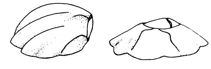
Fig. 13. Comparison of bent (left) and flat (right) barnacle shell morphologies (Lively, 2021).
The barnacle thus grows bent over, and the predatory snails have a much harder time accessing their prey through the opercular plate. This evolution, however, has not been apparent in all individuals of the species. As such, both the unprotected and protected individuals are found in the Gulf of California. The reason for this is twofold: the bent shell takes longer to grow and it makes the barnacle less fecund (Lively, 2021), both of which are direct consequences of the structure of the shell. The shell achieves its bent structure by stopping growth on one side, while the other continues to grow upright. Growing from only one side thus reduces the overall rate of shell development and causes the shell itself to be smaller. In turn, there is less space to house larvae, which makes the barnacle less fecund. While barnacles’ shells can serve to protect them from predation, there are costs to this evolutionary adaptation, which results in the fact that not all individuals can carry this trait.
Limpet Shells
Limpets’ reliance on shell structure to resist being displaced by tidal currents and waves is more important than in barnacles. It is thus logical to think that limpets living in wave-exposed environments will own a shell that is more resistant to wave forces than those living in more sheltered areas. Researchers studied the Fissurella latimarginata species, which is found in both types of environments. Running a multivariate analysis on their data, they found significant differences in shell morphology between F. latimarginata in wave-exposed versus sheltered areas. In exposed environments, limpets typically possess a flatter, elongated shell structure compared to the peaked and round structure found in limpets from sheltered areas (Vasconcelos et al., 2021). As the shell of the limpet grows larger, there is space for a larger aperture, as seen in Figure 14. Thus, the limpet can accommodate a larger foot, resulting in better binding to the substrate (Vasconcelos et al., 2021).

Fig. 14. Relationship between length of the shell (Ls) and area of the foot (Af). Limpets from exposed environments are in black and those from sheltered environments, in red (Vasconcelos et al., 2021).
The limpets with elongated shells and larger feet were found to be able to withstand lab-induced waves of higher velocity. For this species, this variation in shell morphology appears to be an effective strategy in resisting displacement by tidal currents and wave forces.
Much like barnacles, the shells of limpets must also be able to resist impact forces and be resistant to fractures. It has been found that limpets whose shells structural integrity becomes compromised by ocean debris such as rocks, ice, or other objects during storms have a very low survival rate. Once the shell becomes fractured beyond a critical point, the limpet is vulnerable to attacks by predators or may die by dehydration (O’Neill et al., 2018; Taylor, 2016). The shell of the limpet is similar to that of the barnacle in that it is primarily composed of calcium carbonate. However, unlike barnacles, it is believed that the microstructure of the limpet shell may be a contributing factor to its toughness (O’Neill et al., 2018). Researchers conducted experiments to measure impact resistance and fracture toughness of limpet shells and compared their results against the known literature values for calcium carbonate in its mineral form. It was found that the toughness of limpet shells was more than 10 times higher than that of pure calcium carbonate, leading to the hypothesis that this may be directly related to the shell’s microstructure (Taylor, 2016).
Although further research is needed to clarify the relationship between the two factors, preliminary findings have been identified. As seen in Figure 15, the macroscopic surface of the shell is rough. This suggests that crack deflection could contribute to the shell’s toughness. From the initial point of contact, cracks form and propagate. Upon encountering rough edges, they deviate from their original path, which slows their propagation. In doing so, more energy is absorbed and the overall toughness of the material is increased (Faber & Evans, 1983). In fact, research suggests that cracks in limpet shells tend to propagate in the circumferential direction. During testing, the shells show a remarkable resistance to failure in this direction, but the mechanism remains unclear.(O’Neill et al., 2018). Although the specific features of the limpet shell contributing to its toughness remain to be uncovered, the limpet still displays remarkable evolutionary traits to protect itself from common causes of damage in its natural environment.

Fig. 15. View of a limpet shell under scanning electron microscope. Macroscopic roughness is seen in (a); more detailed roughness and cleavages in individual crystals can be seen upon further magnification (b) (O’Neill et al., 2018).
Feeding Mechanisms
Cirri in Action
Barnacles come across yet another critical challenge: how to capture enough nutrients in a shifting marine environment with turbulent waters and irregular currents. Due to their sessile nature, these organisms have a harder time when the water flow is low. Despite the water's limited movement, they must find a way to filter and search for nutrients and plankton.
To address this challenge, barnacles rely on their mobile “teeth”: specialized feeding tentacles named cirri represented in Figure 16.

Fig. 16. Overview of the structure of the common limpet tooth the Patella vulgata (Barber et al., 2015).
The cirri, located at the organism's aperture, rapidly expand and contract in rhythmic, undulating patterns to capture microorganisms (Figure 17).This constant motion is attributable to the collective effort between muscular and cutaneous (skin related) structures (Hindenberg et al., 2022).

Fig. 17. Amphibalanus improvisus's sequential cirral fan movements caught at 500 frames per second. The high frame rate enables precise observation of the fast and complex movements of barnacle cirri. (Hindenberg et al., 2022).
A cirral net or fan is made up of specialized thoracic appendages that are covered by rows of stiff structures resembling a hair or a bristle named setae. It is used by barnacles to seize their prey. As the cirral fan swirls through the water, food fragments are gathered. As presented in Figure 18, a strong water flow, called feeding current, is created during the power stroke by the cirral fan’s movements. Food particles get transported into the “capture zone." During the final stage of the recovery stroke, a vortex is believed to emerge above the barnacle and the food particles are transported by this vortex into the capture zone, where they could be caught during the next power stroke (Trager et al., 1990).

Fig. 18. Balanus crenatus feeding behavior, with the circular fan outstretched to seize food and the red arrow pointing in the direction of the power stroke (Maar et al., 2024).
Moreover, hydrodynamic pressure impacts the marine world by transforming species interactions, which modifies all kinds of life and has the capacity to serve as the main shaping force (Reustle et al., 2023). For example, the ocean’s hydrostatic pressure, which rises by 1 atm per 10m, interferes with a variety of cellular functions: it stiffens membranes and disrupts the ligand binding and protein folding (Ph, 2020). Moreover, wind and the movement of coastal waters stimulate the transfer of sediments and hence influence the evolution of the sea bed (Winter, 2018). Consequently, organisms living in wave-swept settings must adapt their morphological and behavioral traits in order to cope with the hydrodynamic forces that trigger lift, drag, and acceleration (Denny, 1985).
When exposed to water flow, barnacles showed phenotypic versatility in their cirri’s size (Reustle et al., 2023) as well as an alteration of their dietary behavior (Trager et al., 1990). There are three common feeding behaviors that are usually demonstrated by barnacles (Crisp et al., 1997). The first being the “normal beat," when the cirri are extended and dispersed, it expands and then withdraws without a pause. The second, known as the “fast beat," is characterized by the cirri brushing the surrounding water strongly and rhythmically along with partial retractions. Lastly, the “prolonged beat" in which the cirri are spread out and anchored for longer than the other feeding modes. Barnacles exhibit the ability to alternate between active and passive feeding, maximizing their nutritional intake based on their surrounding conditions (LaBarbera, 1981). While active refers to barnacles beating their cirri, passive indicates that the cirri are extended. Usually, most barnacle species beat their cirri during low velocity and extend them at higher current speed (Trager et al., 1990). This implies that their feeding mechanism depends on the water velocity. Furthermore, the passive feeding technique is often more successful and efficient because of its higher capture frequency and reduced consumption of energy.
Additionally, it can be determined, or at least predicted, whether a flow will be turbulent or stable using the handy parameter known as the Reynolds number.

where η is the dynamic viscosity, μ is the flow speed, L is a characteristic length connected to the flow, and ρ is the fluid density.
The dimensionless number is also often expressed as:

where ν = η/ρ is the kinematic viscosity (Arfken et al., 1984).
Appendages comparable to sieves (expanded and spread-out cirri) can act like paddles (the cirri move through the water in a sweeping or paddling motion) at Re < 1, where seawater fluidity becomes more significant than resistance (Cheer & Koehl, 1987). Both the viscous and inertia forces equalize as Re approaches 1. Here, the width of the fluid stream that the filter device encounters is effectively increased by streamline pressure around the cylindrical appendage, consequently raising the rate at which food particles come into contact (Shimeta & Jumars, 1991).
It is important to note that barnacles are known to have smaller, stockier cirri with shortened setae in high-flow locations and longer, thinner cirri with more setae in low-flow places. These organisms’ behaviors react to flow direction shifts instantly, even seeming to sometimes predict fluctuating flow (Trager et al., 1990).
Thus, because of their feeding tentacles, barnacles can adapt to the difficulties of obtaining nutrients in a changing marine environment. The barnacles' feeding process is dependent on water velocity, with distinct feeding patterns observed at different flow rates.
The Feeding Power of Limpet Radula
Limpets have the challenging task of consuming algae that accumulates on rocky terrain. To deal with this challenge, limpets developed a biological mechanism that can survive the daily damage and friction caused by scratching the rough surfaces. Lopese tails, or radulas, are tongue-like appendages coated with tiny teeth that limpets use to scratch paths in the rocks while feeding on phytoplankton. The tracks reveal how strong those teeth are, but scientists did not quantify it until 2015 (Skazál, 2023).
The feeding organ of the limpet, the radula, has nearly 100 rows of teeth. As shown in Figure 19, the teeth develop and move forward along the radula as they mature. While the more basic doccoglossan radulae work by pulverizing stony surfaces and using their teeth as shovels to scrape and crush algae out of them, the rhipidoglossan radulae are employed as rakes or brooms to brush and gather marine debris, as illustrated in Figure 20. The teeth deteriorate when they start to work, usually one tooth row per day. After that, a new row of teeth erupts to replace the worn-out ones, ensuring that the limpet can continue to effectively graze on hard surfaces. (Ukmar-Godec et al., 2015).

Fig. 19. In motion, limpet radula with a blue arrow indicating the direction of stroke, (c) an optical micrograph of the mature end, (b) an extracted radula. (Ukmar-Godec et al., 2015).

Fig. 20. Diagram showing two varieties of herbivorous limpet radulas: rhipidoglossane (right) and doccoglossane (left), where R stands for center tooth and MT for marginal teeth, LT for lateral teeth, mLT for minor lateral teeth, and MLT for major lateral teeth (Ukmar-Godec et al., 2015).
It is important to mention the tensile stress σ, a force distribution per unit area along the applied load’s axis: (Morrow & Kokernak, 2011).

Furthermore, the main component of limpet teeth is made up of goethite nanofibers, making up around 80% of their volume. These goethite nanofibers are typically many micrometers in length but just a couple tens of nanometers in diameter, which is below the size threshold recognized as promoting flaw insensitivity (Barber et al., 2015). Thus, the resilience of the mineral nanofibers in the composite structure must be a crucial factor in the limpet tooth’s strength. While the teeth experience shear stress during grinding, it has been proven that the density of defects in the reinforcing phase determines the tensile strength of manufactured composite materials that include fibers with diameters more than two orders of magnitude greater than those of nanofibers detected in limpet teeth. Hence, compared to bigger fibers, limpet teeth can withstand flaws better. Because of their special structure, the radulae could potentially be used to incorporate nanofiber components below a critical size that characterizes defect tolerance, thereby optimizing composite toughness (Barber et al., 2015).

Fig. 21. Images captured by a scanning electron microscope that show how a limpet tooth sample was evaluated mechanically. (a) The limpet tooth is placed in the AFM cantilever setup with a portion secured by gripping glue. (b) After the tensile test, the fracture point, which highlights the tooth’s failure characteristics, is visible at the sample free length mid-point (Barber et al., 2015).
In addition, for the purpose of determining the material’s failure behavior, shown in Figure 21, and explaining the failure of the reinforcing mineral phase in the tooth, the limpet tooth’s failure was initially assessed using a macroscopic fragmentation test (Barber et al., 2015). As seen in Figure 22(a), limpet teeth were embedded in solid epoxy resin and prepared for tensile testing. Using backscattered electron imaging on a polished surface of the epoxy-embedded limpet tooth, it was possible to observe the nanofibrous goethite. Tensile testing of hydrated specimens showed that a shorter average nanofiber length correlated with higher tensile strain, as illustrated in Figure 22 (b). This behavior reflects a gradual fragmentation of the reinforcing phase, rather than interfacial debonding or fiber pull-out—mechanisms typically observed in other tough biological composites. Consistent with composite failure theory, the nanofibers undergo splitting and length reduction until a stress saturation plateau is reached. These findings suggest that stress is effectively transferred within the tooth structure, generating sufficient tensile load on the goethite nanofibers to induce their failure. In tensile testing, the mechanical integrity of the nanofibers is directly impacted by the ensuing tensile stress. This can lead to failure when these fibers are overstressed beyond their tensile strength. Therefore, the limpet tooth’s potential strength is defined by the mineral phase failure (Barber et al., 2015).

Fig. 22. The structure of limpet teeth was examined for failure by (a) embedding the teeth in epoxy resin and applying tensile stress, (b) plotting the length of the nanofiber during straining, which demonstrated fragmentation upon failure (Barber et al., 2015).
Tensile tests on limpet teeth revealed a relationship between their structure and mechanical performance. All specimens exhibited a linear elastic behavior; however, as shown in Figure 22(b), nonlinearity was evident when the sample strain surpassed 2%. Due to the lack of a highly ordered reinforcing phase distribution within the limpet tooth, variability in the stress-strain curves appears. For example, if the discrete volume under test had a comparatively larger proportion of mineral phase than other samples, a greater elastic modulus might be anticipated (Barber et al., 2015).
The relevant structure that was evaluated can be used to explain the tooth’s deformation. Since the mechanical characteristics of the reinforcing mineral are anticipated to take-over the stress-strain reaction, limpet teeth have a large volume proportion of mineral phase. The nonlinear stress-strain behavior may be influenced by the weaker protein matrix, particularly because the tooth’s goethite phase has been demonstrated to be linearly elastic. Nonetheless, the tensile tested limpet tooth samples from the plot in Figure 23 has a linear elastic modulus of 120 ± 30 GPa, which is higher than the projected polymeric value and closer to the 180 GPa observed for the pure mineral phase (Barber et al., 2015). Therefore, it is justified that the mineral phase dominates the deformation behavior of limpet teeth. Figure 23’s maximum tensile stress at limpet tooth sample failure illustrates the dataset’s variety. The degree of variation in the limpet tooth material’s strength can be attributed to two factors: the defect insensitivity of the mineral nanofibers or the length of the sample tested, as indicated by assessments of stress concentrations surrounding flaws (Barber et al., 2015).

Fig. 23. Individual limpet tooth samples of different lengths were evaluated using AFM to determine the stress-strain behavior till failure (Barber et al., 2015).
Ultimately, limpet teeth exhibit extraordinary tensile strength, driven by their minerals. Their failure is dominated by nanofiber fragmentation, making them one of the strongest natural materials.
Conclusion
By studying the physics behind barnacles and limpets’ survival mechanisms, one understands how nature has developed design solutions to help them withstand environmental stresses and survive in their habitats. The ubiquitous environmental stress that limpets face due to survival in intertidal zones is wave pressure and water turbulence. These stresses pose difficulty to move without losing adherence to the substrate. As a design solution, limpets developed the mechanism of control while crawling through ditaxic contractions and mucus heterolysis. In addition, exposure to the sun entails threats of desiccation and heat stress for limpets, which can consequently affect their mucus effectiveness. To avoid desiccation, limpets developed the habit of moving during nocturnal high tides and returning home before sunrise.
To maintain their sessile lifestyle, adult barnacles must adhere efficiently to their substrate. They endure extreme hydrodynamic forces in their environment and counter these by releasing a high-strength adhesive that permanently anchors them to the surface. Moreover, since barnacle substrates can be versatile, barnacles developed a design solution to account for the nature of the substrate. Barnacles secrete different amounts of cement depending on the substrate’s polymeric substances, which helps them adapt to different environments.
In addition, barnacle shells help resist mechanical stresses imposed by tidal waves. However, predation pressure also presents a grave threat to barnacle populations, as they can be pried off their substrate by predators, like snails. In these cases, not only do shells resist hydrodynamic pressures, but they also evolve as a defensive adaptation. Barnacle shells develop in different shapes and angles. This specialized shell morphology stands as a design solution for barnacle predators.
Lastly, in intertidal zones, limpets face the environmental stressor of feeding on hard, rocky surfaces. They must crape algae from rough substrates and endure the severe circumstances of low tide desiccation. These challenges make it difficult for limpets to gather food efficiently while avoiding damage to their feeding mechanism or moisture loss. To counter these problems, limpets evolved a radula, a unique structure composed of rows of powerful teeth. Additionally, limpet’s radula regenerate continuously, allowing them to maintain their feeding effectiveness over time, despite the wear caused by scraping rocks.
Indeed, barnacles and limpets exemplify nature’s brilliance in evolving design solutions to external stresses, making them valuable models for studying sustainability across numerous engineering fields.
References
References
Anderson, D. T. (1993). Barnacles: Structure, function, development and evolution. Springer Science & Business Media.
Morrow, H. W., & Kokernak, R. P. (2011). Statics and strength of materials (7. ed). Prentice Hall.
Southward, A. J. (Ed.). (2018). Barnacle Biology. Routledge. https://doi.org/10.1201/9781315138053