
Table of Contents
Keywords: Yew, Taxus baccata, alkaloids, taxine, paclitaxel, toxicity, adaptation, photosynthesis, structural defenses, ecological interactions
Abstract
It is undeniable that the yew tree, Taxus baccata, exhibits an exceptional chemical profile. With its many defensive secondary metabolites, it has evolved potent mechanisms that enable it to withstand numerous environmental challenges and threats surrounding it, like pathogens or abiotic stress. It has evolved to adapt its leaf structure and photosynthesis in accordance with the quantity and quality of light it receives. Then, the specificity of its toxicity may relate to the tree’s strategy for protection during its early, vulnerable stages — preventing consumption by insects or disease — since it grows extremely slowly and takes time to become strong and robust. Simultaneously, this selective toxicity allows it to form a mutualistic relationship with birds who will spread its seeds. Thus, the yew tree stands as a pertinent example of the way plants balance defense and survival with ecological interactions.
Introduction
Only 1 (one) lb (0.45 kg) of yew clippings is needed to kill a 1,000 lb (450 kg) animal. Although yew hedge clippings can occasionally seem fresh and alluring to calves, data indicates that even a tiny quantity of leaf or seed material can quickly kill cattle (Gower, 2006). This startling discovery demonstrates just how strong the tree's chemical defenses are: yews produce strong secondary metabolites, particularly the taxine alkaloids, which impair heart function and can cause abrupt collapse. The yew developed these diverse biochemical defenses as an effective deterrent against grazing, as a barrier against microbial attack and decay because of the constant pressure from herbivores, harsh weather, and competition for light and nutrients. Consequently, the plant becomes both ecologically durable and extremely harmful to any animal that eats its leaves or crushes its seeds.
On a broader scale, the yew tree (Taxus Baccata) is one of the oldest evergreen conifers in the Taxaceae family, and is well-known for its durability, longevity, and impressive look. Yews, which are native to Europe, North Africa, western Asia, and portions of East Asia and North America, do well in temperate areas and frequently like well-drained soils, churchyards, and shaded forests (Thomas & Polwart, 2003).
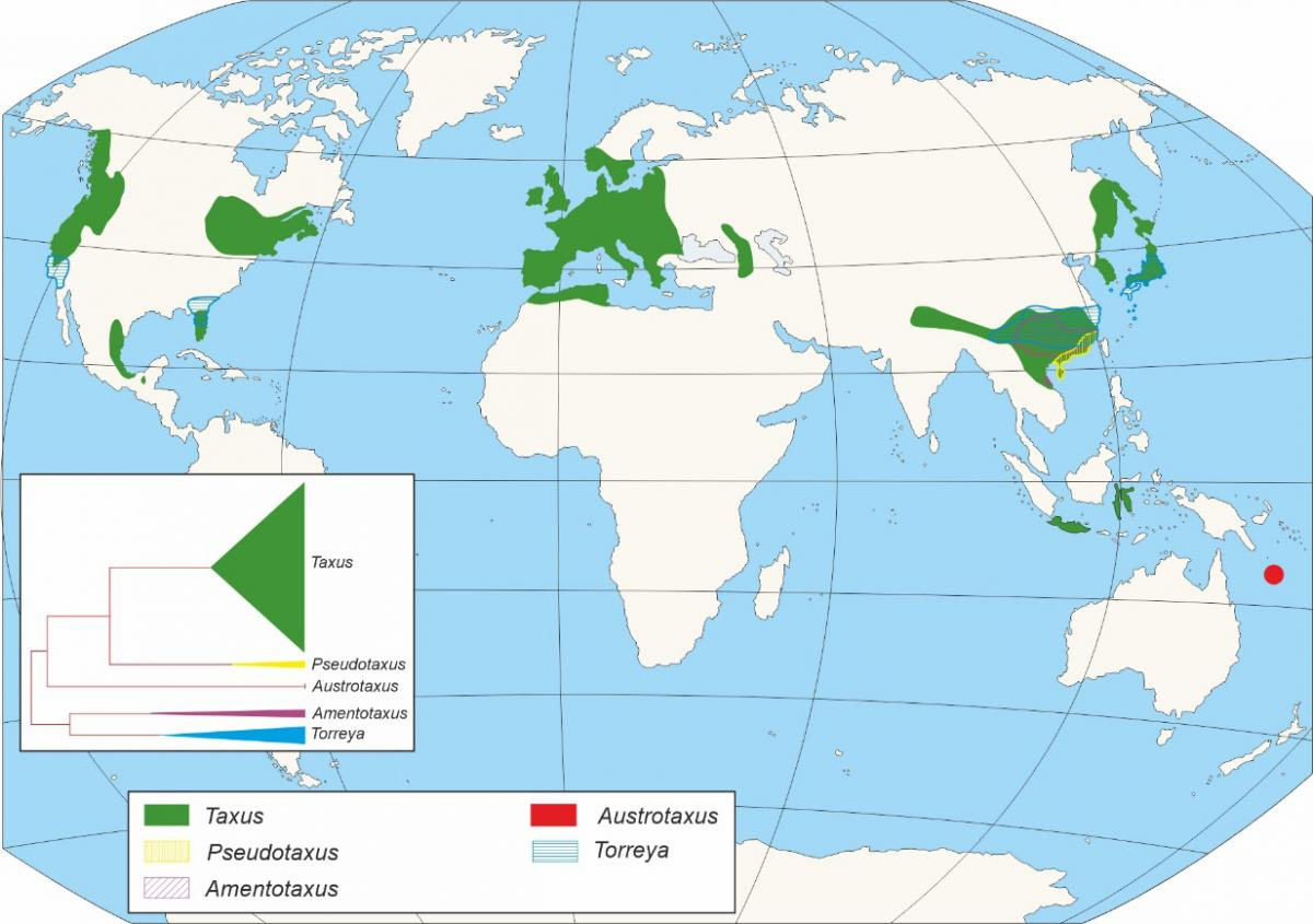
Fig. 1. Distribution of Taxus Baccata in the world (History of Yew Forests in Northern Iberian Peninsula | LIFE Baccata, 2025).
In churchyards and cemeteries, especially, yew trees have long held cultural and spiritual significance as symbols of immortality, endurance, and the cycle of life and death. The yew is a visually striking and ecologically interesting tree, distinguished by its vivid red arils encasing hard seeds, peeling reddish-brown bark, and flat, dark green needles set in neat rows along its branches (History of Yew Forests in Northern Iberian Peninsula | LIFE Baccata, 2025).

Fig. 2. English Yew (Max, Plantura Garden, 2023).
The species is known for having some of the longest lifespans in the world; some of these trees have been seen to survive for over a thousand years, and other old trees, like the Fortingall Yew in Scotland, are said to be over 4,000 years old (Hageneder, 2013). With its complex chemical profile, the yew raises questions regarding the molecular structures that make up its toxic components and how these have shaped its ability to defend itself specifically against threats and to grow in challenging environments.
Yew Needles and Photosynthesis
Yew Needle Structure and Function
The yew tree’s leaves are small, flattened needles with a pointed tip as shown in Figure 3. The needles are dark green in color and grow in rows on either side of each twig. Unlike broadleaf trees, such as maple or oak, the yew tree’s needles are thick, waxy, and long-lived. Beneath the protective waxy layer lies the mesophyll, a dense tissue rich in chloroplasts. Photosynthesis is carried out entirely within a yew’s needles, in the chloroplasts.

Fig. 3. Yew (Taxus Baccata) needles (Yew - Tree Guide UKK, n.d)
Light plays a direct role in controlling plant growth and development by facilitating photosynthesis and triggering a range of photomorphogenic responses, the responses of organisms to light signals that regulate changes in structure and form (Devaney et al., 2014). Growth and survival of plants is determined by the quantity and quality of light. The question is, how does the yew tree chemically adapt its needles to survive, photosynthesize, and protect itself across light extremes?
Photosynthesis and the Light Reactions
Photosynthesis is a biochemical process in which sunlight is harvested to synthesize glucose from carbon dioxide and water, illustrated in Equation 1 (Cooper, 2012).
6CO2 + 6H2O + light → C6H12O6 + 6O2 (1)
Through converting light energy into chemical energy, photosynthesis is a primary source of metabolic energy for photosynthetic organisms, including plants, algae, and some bacteria. This process takes place in two distinct stages: light-dependent and light-independent reactions (also known as the Calvin cycle).
In light-dependent reactions, energy from sunlight drives the synthesis of ATP and NADPH products, as well as the formation of O2 as a byproduct from water splitting. For this process, sunlight is absorbed by photosynthetic pigments, known as chlorophylls in plants. These are organized into photocenters in the thylakoid membrane, photosystems I and II. These systems absorb photons and transfer their excitation energy to a reaction center chlorophyll (Fig. 4) (Cooper, 2012).

Fig. 4. Simplified representation of the light-dependent reactions showing Photosystem II and Photosystem I (BioNinja, n.d.)
Excess light, Photoinhibition and Photoprotective Mechanisms
Within the light reaction, photosystem II (PSII) plays a particularly important role. Counterintuitively, PSII is the first complex in the electron transport chain and is responsible for the oxidation of water, resulting in the release of O2 among other products. Since PSII directly absorbs photons and catalyzes this reaction, it is most susceptible and vulnerable to damage due to environmental light extremes. If absorption of photons exceeds what can be processed in the later reactions, the excess excitation energy can accumulate in the chlorophyll molecules, resulting in a decline of PSII activity, a phenomenon known as photoinhibition. This will reduce the plant’s overall photosynthetic capacity. At the molecular level, excess excitation energy in chlorophyll molecules can transfer to oxygen, forming reactive oxygen species (ROS) such as singlet oxygen and superoxide radicals (Li et al., 2023). These are highly reactive molecules which oxidize proteins, lipids, and pigments within the thylakoid membrane, worsening the effects of photoinhibition.
To test this, an experiment by Devaney and his colleagues was conducted in which plants were divided into three light environments: low light (<5% full daylight), medium light (5-20%), high light (>20%) (Devaney et al., 2014). After some time, the needles were analyzed for photosynthetic efficiency using chlorophyll fluorescence, a non-destructive measurement of PSII activity. Numerous factors were measured, including the maximum quantum efficiency of PSII, effective quantum yield (how much absorbed light energy was used in photosynthesis), and electron transport rate (how quickly electrons move through PSII). These parameters altogether exhibit whether needles are efficient, promoting healthy photosynthesis, or stressed, resulting in light damage (Devaney et al., 2014).
The results of this experiment showed that in bright, intense light, photosynthesis efficiency drops. As shown in Figure 5, the maximum quantum yield of PSII (FV/FM) decreases as light intensity increases. Although efficiency dropped, the effective quantum yield and electron transport rate were both found to be optimized for low intensity light in shaded plants, which is evidence of shade acclimation (Devaney et al., 2014).

Fig. 5. Comparison of growth light-intensity–dependent changes in the maximum quantum yield of PSII (FV/FM) (Wu et al., 2020).
Although the yew is normally an understory species adapted to low light, its needles can still face sudden bursts of intense light, known as sun flecks, when light penetrates through canopy gaps (Durand et al., 2022). This still puts the photosynthetic system at risk of photoinhibition. The solution to how Yew needles protect their photosynthetic machinery from damage due to excess light intensity lies in the activation of several chemical and structural defense systems. These include non-photochemical quenching, the xanthophyll cycle, and epicuticular wax.
The first chemical defense deployed is non-photochemical quenching (NPQ), a mechanism in which excited chlorophylls quickly dissipate their energy into harmless heat inside the pigments of PSII (Li et al., 2023). Instead of allowing excess photons to drive dangerous reactions, this way, energy is efficiently dissipated as molecular vibrations. This process is known as the fastest and most efficient response of the photosynthetic membrane to excess light (Zuo, 2025).
Next, the xanthophyll cycle is another chemical defense that the system uses to prevent photodamage. Carotenoids are accessory pigments in plants which produce yellow, orange, and red colors and play an essential role in photoprotection (Linus Pauling Institute, Oregon State University, 2019). There are three carotenoid pigments that are involved in the xanthophyll cycle: Violaxanthin, antheraxanthin, and zeaxanthin (Li et al., 2023). Violaxanthin, a pigment which is most present in low light conditions, known as the light-harvesting form, has two epoxy groups (-O- bridges). Antheraxanthin, the pigment found in medium light conditions, has one epoxy group. Zeaxanthin has zero epoxy groups, and is the heat-dissipating form, as it is most present in high light conditions. Under intense light, the enzyme violaxanthin De-epoxidase (VDE) removes epoxy groups from the pigment violaxanthin, converting it sequentially into antheraxanthin and zeaxanthin. When light drops, enzyme zeaxanthin epoxidase (ZE) reverses this process, made visually clear in Figure 6. This cycle is critical for balancing energy dissipation and absorption, ensuring excess energy is safely released rather than causing damage (Li et al., 2000).

Fig. 6. The xanthophyll cycle showing enzymatic conversion of violaxanthin to antheraxanthin and zeaxanthin under high light via VDE, and the reverse conversion under low light via ZE (Li et al., 2000).
A third defense used is the surface chemistry of the needle, specifically its epicuticular wax layer. The wax is a hydrophobic layer of long-chain alkanes, esters, and alcohols that scatter and reflect light (Li et al., 2023). This scattering lowers the photon flux that reaches the chloroplasts. With this chemical defense comes a trade-off, where although there’s more physical protection from intense light, the wax layer slightly limits CO2 diffusion, lowering gas exchange efficiency.
Low Light and Pigment Adaptation
While these chemical defenses protect the yew from light excess, most of its life unfolds under the opposite condition, in constant shade. Yew trees are commonly found in the lower canopy beneath larger trees, receiving <2-3% of light (Devaney et al., 2014). In coniferous forests, optical filtering by leaves occurs where light passing through a canopy is spectrally filtered (Rautiainen et al., 2018). Different wavelengths are selectively absorbed and scattered as light travels downward through a forest. Under deep shade, light available to the yew tree is much dimmer and shifted in color, mainly green/blue wavelengths as red light has been absorbed by the upper canopy leaves. Yew needles, like all leaves, contain two main forms of chlorophyll, a and b. Chlorophyll a is the primary pigment which directly drives photosynthesis by transferring energy to reaction centers. Chlorophyll b is a supporting pigment, typically found in lower quantities, which broadens the spectrum of light that can be absorbed. At low levels of light, the yew adapts by increasing its chlorophyll b relative to chlorophyll a, lowering the ratio between them. Therefore, allowing the plant to capture more of the wavelengths that reach it in low light.
Ecological studies of natural yew populations were conducted and demonstrate this chemical adaptation. This study analyzed how the occurrence of yew seedlings and saplings correlate with light availability, measured by photon flux density (PPFD) (Iszkuło & Boratyński, 2005). In simpler terms, how does the amount of usable light reaching the forest floor influence where yews can germinate, survive, and grow? It was found that very young seedlings (<6 cm) were found primarily in highly shaded plots, meaning a low PPFD. While intermediate seedlings (6–25 cm) and larger seedlings (25-100 cm) thrived in moderate light levels between 2–7 % of full daylight. At higher light intensities (PPFD>7 %), seedling density declined sharply as yews were outcompeted by faster-growing plants. These findings suggest that yew seedlings are biochemically and ecologically optimized for life in the understory, relying on shade-acclimated photosynthetic machinery for survival (Iszkuło & Boratyński, 2005). Yet even as they quietly thrive in low light, yews carry an inherent chemical arsenal that quietly ensures their protection; a trait that will later reveal itself in their deadly reputation.
One Bite of Japanese Yew Can Kill a Cow
The yew tree has long been associated with graveyards and death. For instance, in Shakespeare’s Twelfth Night, Viola’s lament, it says “I am slain by a fair cruel maid. / My shoulder of white, stuck all with yew” (2.4.60). This depicts the yew’s connection to death and mourning. In fact, Laertes killed Hamlet with a blade poisoned with yew. Moreover, with its remarkable longevity, this tree symbolizes a testament to nature’s resilience. This ability is in part due to its toxic properties. Though nearly all plants contain toxins and carcinogens, a single bite of the Japanese yew can kill a cow (Stegelmeier & Davis, 2023). This poisonous superpower is a defense mechanism that deters herbivores and protects the conifer from being consumed by wildlife; it allows the yew to endure across generations.
Taxines
This tree of death contains a heterogeneous blend of cardiotoxic alkaloids. This means it contains a nitrogenous organic compound that can harm the heart. The first investigation of the deathly effects of the English yew (T. baccata) occurred in the 19th century, when a scientist named Lucas phytochemically analyzed the leaves and recovered a white, non-crystalline powder. Then, a French scientist, Marmé, isolated a crystalline form of the taxine. At the end of the 19th century, Hilger and Brande derived a basic molecular formula for taxine: C37H52NO10 (Wilson et al., 2001).
The two prominent and most cardiotoxic alkaloids isolated from this plant are Taxine A and Taxine B, though there are more than a dozen unique alkaloids (Handeland et al. 2017).
About 1.3% of the alkaloid isolated from the leaves of the English Yew was Taxine A. Its molecular formula, as shown in Figure 7, was revealed using spectrophotometric analysis, a technique that measures how compounds absorb light at specific wavelengths to determine their chemical structure and concentration. A greater fraction of the extracted alkaloid came from Taxine B, representing an astonishing 30%. This taxine’s chemical formula is visualized in Figure 8.

Fig. 7. Taxine A; 2α,13α-Diacetoxy-7β,10β-dihydroxy-9-oxo-2(3→20)-abeotaxa-4(20),11-dien-5α-yl (2R,3S)-3-(dimethylamino)-2-hydroxy-3-phenylpropanoate; or C35H47NO10 (“Taxine Alkaloids”).

Fig. 8. Taxine B; 10β-Acetoxy-1,2α,9α-trihydroxy-13-oxotaxa-4(20),11-dien-5α-yl (3R)-3-(dimethylamino)-3-phenylpropanoate; or C33H45NO8 (“Taxine Alkaloids”).
From the analysis of the compounds and their molecular formulas, important chemical characteristics of taxines have been identified. For instance, they are unstable in a neutral or alkaline environment. However, they are heat-stable, therefore, drying or boiling them does not reduce their toxicity (Labossiere Thompson, 2018). As such, their stability increases, maintaining the deadliness, if they are converted into sulphate salts. (Wilson et al., 2001).
Of the two, taxine B along with its derivative have stronger cardiac effects (Handeland et al. 2017). These compounds mainly affect the heart and have minor effects in other organ systems (Wilson et al., 2001). By researching the effects of the taxines on cardiac myocytes and medullated axons, it was discovered that they cause an increase in cytoplasmic calcium concentrations which alter the calcium and sodium ion channel conductance (Lee et al., 2024). In fact, the taxines were later discovered to be calcium and sodium channel antagonists. As shown in Figure 9, which is a diagram used to represent the ion channels of the phospholipid bilayer, this causes them to decrease Na+ and Ca2+ influxes and block the electrical signals needed for coordinated heartbeats. Then, this respectively slows depolarization and weakens cardiac contractions (Wilson et al. 2001).

Fig. 9. The cardiac myocyte. Taxine B primarily inhibits fast sodium channels and L-type calcium channels in cardiac myocytes (Pettengill, 2022).
As such, taxine B induces a negative inotropic effect, while increasing the A-V conduction time and QRS duration. The increase of two terms respectively means that the blockage of the sodium and calcium ion channels weakens the muscle contraction of the heart while increasing the time it takes for electrical pulses to travel through the heart and for the ventricles to contract during a heartbeat. Adversely, taxine A only mildly reduces heart rate (Wilson et al., 2001).
These effects can be recognized by cardiac arrhythmia, atrioventricular block, and diastolic cardiac arrest in patients who have ingested the toxin. This often leads to acute heart failure within 24 hours of ingestion (Handeland et al., 2017). In fact, the cardiotoxic taxine alkaloids are present in all parts of the tree other than the fleshy aril that surrounds the seed that is shown in Figure 2 in the intro.
Variability of Toxicity
The toxicity of the yew varies according to its plant parts, its species, and through the seasons. A study conducted in 2002 shows that the stems of the yew are more toxic than the needles, which correspond to the leaves of the yew, shown in the figure above (Shanker et al., 2002). The English Yew and the Japanese Yew report the most abundant concentration of taxines A and B of the Taxus genus, while the Pacific Yew contain only trace amounts of these alkaloids (Lee et al., 2024). Maximal concentrations of the taxines occur in the winter, though it is seemingly consistent through drought periods (Wilson et al., 2001).
The yew tree has justifiably earned its reputation as the “tree of death”. The previously mentioned statement “one bite of Japanese Yew can kill a cow” serves as a stark testament to its lethal potential (Stegelmeier & Davis, 2023). The dosage needed to kill other animals and human is similarly low. Humans are highly sensitive to taxine poisoning with only 0.6 to 1.3 g of the plant per kg of body weight, equivalent to 3 to 6.5 mg/kg of taxine being a lethal dose, which is equivalent to a few leaves. Due to its fat-soluble structure and large molecular size, it is also quite difficult to remove from the body (Labossiere & Thompson, 2018). It is only toxic by ingestion, not by touch. If a person were to ingest any deadly part of the yew, there would not be an antidote for this poisoning. The treatment would include supportive care like cardiopulmonary resuscitation or extracorporeal life support (ELCS/ECMO) in cases of severe ingestion (Labossiere & Thompson, 2018).
Selective Toxicity
The development of toxic foliage, bark, and seeds represents a key evolutionary adaptation that significantly contributes to the longevity of the yew tree by deterring herbivory. There are many cases where animals have been found dead near yew trees with remnants found in their rumen. Such is the case for free-ranging ruminant in Norway 2008, where a group of animals were found intoxicated and deceased within a garden of yew plants (Wilson et al., 2001). However, anecdotally, many have reported some animals to be tolerant to the yew. The antelope, deer and elk may be able to ingest yew without further ramifications attributable to ruminal detoxifications of the taxines (Lee et al., 2024; Wilson et al., 2001). These species will generally only forage this plant if they must, for example when it is the predominant food source or during the winter when other food sources are scarce (Wilson et al., 2001). As such, the yew can live a very long time and does not need to fear being eaten as a young tree. Moreover, by minimizing damage from animals, the yew conserves metabolic resources that would otherwise be required for tissue regeneration, thereby supporting slow and sustained growth, structural stability, and long-term survival. This is a common strategy in plants that cannot physically escape a herbivore. Instead, they deter predators by accumulating poison. This is a necessary evolutionary trait for yews, a species of slowly growing plants that stay young and therefore attractive for herbivores for a long period of time.
The resourceful yew has found multiple ways to use the arils as an evolutionary mechanism for their survival and reproduction. Though the tree is toxic, it needs to find a way to disperse its seeds. Many trees will form symbiotic relationships with birds, insects, or pollinators. How could yew achieve this if all parts of it were toxic? As an evolutionary adaptation, the yew seeds are noxious, but the arils themselves are edible. They are reported to taste like honey when ripe and serve as high caloric snacks for birds and animals during the fall and winter (Scotland’s Yew Tree Heritage Initiative., 2021). The yew first attracts birds with its bright, mostly red, arils, whose color can be attributed to retro-carotenoid rhodoxanthin (Schex et al., 2021).
Then, the digestive track of birds has a symbiotic relationship with the toxic design of yews. Thrushes, like blackbirds, redwings, and fieldfares, eat the whole aril. Unlike mammals, they do not chew or crush the seeds which would release taxines into the gut and poison the animals. The avian gastrointestinal (GI) tract (Figure 10) has adapted to digest and absorb nutrients for their fast metabolism quickly and efficiently. Birds in the Turdidae family have thick epithelial linings to protect them when swallowing whole food items. The esophagus travels the food into the crop and into the stomach. The stomach is separated into the glandular stomach (proventriculus) and the muscular stomach (gizzard). The latter grinds the food whereas the proventriculus mixes it with digestive juices. This breaks down the soft aril without damaging the hard seed. The seed passes into the intestine and out the cloaca (Klasing, 1999).

Fig. 10. Internal anatomy of a bird. (All About Bird’s Digestive Systems: From Beak to Tail, 2024).
This aril-seeds strategy keeps the toxic seed intact through avian digestion. These birds can then disperse the seeds far through droppings. The thrush’s migratory behavior favors the spread of the seeds across wide areas (Stanley, 2015) where the seeds are fertilized by the bird’s feces. This mutualistic relationship between birds and yew is an ingenious design solution that highlights the plants' selective toxicity as an evolutionary strategy for reproduction (Scotland’s Yew Tree Heritage Initiative, 2021). The balance of defense and reproduction allows the yew to thrive in harsh environments, illustrating how natural selection shapes complex, adaptive traits to overcome ecological challenges.
Fighting Tiny Invaders: Yew’s Antifungal Chemistry
Conifers like the yew, spruce, and fir are often plagued by several fungal pathogens such as Heterobasidion (annosus root rot, Figure 11A), Phaeolus schweinitzii (Schweinitzii butt rot), and Perenniporia subacida (stringy butt rot, Figure 11B). These fungi attack weak points in the bark or wood, often close to soil where they are produced and in old injuries like bark cracks or logging scars (Canadian Forest Service, 2024). While these fungal pathogens are relatively non-lethal, they can severely weaken trees by hollowing the major trunks and roots, making the wood susceptible to breakage by wind or infection by more aggressive pathogens such as oomycetes (Callan, 2024).

Fig. 11. (A) Annosus root rot (H. occidentale) on the trunk of a tree (Carpentier, 2024). (B) The damage down by stringy butt rot (P. subacida) inside the trunk, notice the hollow, weakened wood and the mycelium spores in white (Allen, 2011).
Oomycetes, commonly known as water molds, are another common pathogen that yew trees face, which are fungus-like organisms that either live on decayed plants as saprotrophs or survive off other plants as parasitic organisms. Zoospores spread through water and contact roots or trunks of trees or plants, germinating into the host and eventually killing them. Famously, the Irish potato famine was caused by an oomycete, Phytophthora infestans, or Late blight (Editors of Encylopaedia Britannica, 2017). Conifers are faced with infestations and disease caused by oomycetes such as Phytophthora root rot (Missouri Botanical Garden, 2024). This disease causes the feeder roots, which are essential to the tree's growth and development, to rot and obstruct the tree's ability to absorb water. The tree’s needles will turn yellow, and it will eventually die. Another fungal disease that particularly affects yew trees is the Yew Needle Blight (Sphaerulina taxi). It primarily attacks new growth, where infected needles will first turn pale green before changing colors to grey or reddish-brown, after which they are eventually discarded from the shrub (Figure 12). Most of the time, tiny black fruiting bodies will appear on the affected needles, which is a clear sign of infection (Pacholko, 2023).

Fig. 12. A yew tree infected by needle blight (Plant Addicts, 2019).
In the face of these fungal assailants, yew had developed a powerful chemical defense mechanism which they incorporated into their tissues: paclitaxel. Also known as the trade name Taxol, this compound is now perhaps most well-known for its role in cancer treatment. But yew trees have no care for the tumors of humans; instead, this secondary metabolite provides defense against parasitic organisms who wish to multiply on their tissues, by inhibiting the parasite’s cellular division.
Chemical Structure and Mechanism of Paclitaxel
The chemical structure of paclitaxel (C47H51NO14) is vital in its effect against mitotic division. It has a chemical structure that is mostly composed of a taxane ring, a fused tricyclic ring with two cyclohexanes and one cyclooctane, which is the characteristic structure of compounds produced by yew trees such as paclitaxel, baccatin, and docetaxel. Paclitaxel differs from all other taxanes because of its N-benzoylphenylisoserine group, a large, bulky side chain that is attached via an ester linkage to Carbon 13 of the ring composed of benzine, hydroxyl, and amino groups as seen labelled below in Figure 13 (Alves et al., 2018).

Fig. 13. The chemical structure of paclitaxel (Taxol). Notice Carbon 13, marked on the cyclohexane ring, is chiral and attached to a N-benzoylphenylisoserine group, an important sidechain that differentiates paclitaxel from other taxanes (Alves et al., 2018).
Paclitaxel’s famous anti-cancer properties come from its ability to promote mitotic spindle stabilization through inducing the polymerization of microtubules. Microtubules are proteins that are a vital part of the cell. They are not just structural components, but also actors in cell signaling, transport of cell components, and more. Microtubules are fundamental in cell division because they form the spindles fibers that pull sister chromatids apart in anaphase of mitosis, without which cell division cannot occur. Each strand of this cellular component is made up of 13 longitudinally aligned protein fibers which are then composed of several heterodimers of the protein tubulin 𝛼 and 𝛽. This multi-leveled organization allows the microtubule to shorten and elongate by adding tubulin molecules at the ends through GTP catalyzation, an ability named “dynamic instability” (Figure 14). Dynamic instability is particularly important in the mitotic phase as microtubules must reorganize, attach to, position, and separate chromosomes, all of which require constant polymerization and repolymerization. Mitosis is thus interrupted, and cell division does not occur, sometimes even causing apoptosis, or cell death, due to its effects on apoptotic pathways which are not yet fully understood (Alves et al., 2018).

Fig. 14. The structure of microtubules and its polymerization, and depolymerization process. This is an important property of microtubules that gives it its function (Borys et al., 2021).
The inhibition of dynamic instability, either through stabilizing or destabilizing microtubule polymerization, would therefore have catastrophic effects on cellular replication (Alves et al., 2018). Taxanes such as paclitaxel bind to microtubules and cause microtubular dynamic suppression. The exact chemistry of paclitaxel binding to microtubules is not yet completely clear due to paclitaxel’s complex shape and dynamic binding pocket, which makes it difficult to observe with current technology (Bozdaganyan et al., 2025). The three-dimensional shape of the molecule can be described as an “inverted cup-shape”, with the bulky sidechain lying across the “mouth” of the cup as seen in Figure 15 (Kingston, 1991).

Fig. 15. 3D stereochemistry representation of paclitaxel. The taxane group forms a cup-like structure with the sidechain extending out of the cup and curving around like a lid (Kingston, 1991).
The consensus on paclitaxel’s chemical mechanism involves the molecule binding to a specific binding site on the 𝛽-tubulin subunit of the microtubule on the luminal, or inner, side. Research suggests that paclitaxel stabilizes microtubules through a few different mechanisms. First, paclitaxel constrains the M-loop of 𝛽-tubulin, which is a flexible structure that connects different sections of a microtubule, as seen in Figure 17B. The attachment of the compound into the binding pocket stops the M-loop from buckling and moving, restricting the microtubule’s ability to be flexible and hinder dynamic instability (Bozdaganyan et al., 2025). To continue, paclitaxel differs from other taxanes as its bulky side chain recognizes assembled microtubule structures as opposed to unconstructed tubulin conformations. It preferentially binds to “straight tubulin” (Figure 18A), which is observed in assembled microtubules, versus “curved tubulin” (Figure 18B), which is observed in unassembled tubulin, encouraging microtubule stabilization and hindering mitosis (Prota et al., 2023). Furthermore, it lowers the concentration of tubulin required for mitotic spindle assembly, inducing mitosis in unfavorable conditions such as low temperature and absence of required proteins and GTP (Young et al., 1992).

Fig. 17. (A) Tubulin heterodimer (α-tubulin in gray and β-tubulin in white) (B) Tubulin β-subunit structural features. Things to note include the taxane binding site circled in purple, and the M-loop which is adjacent to the taxane binding site (Adapted from Prota et al., 2023).

Fig. 18. Paclitaxel inside the binding pocket of (A) a curved tubulin, observed in unassembled microtubules and (B) a straight tubulin, observed in assembled microtubules, which paclitaxel preferentially binds to (Prota et al., 2023).
Paclitaxel’s Effectiveness Against Pathogens
Experimentally, paclitaxel has been found to be effect against various fungi from different taxonomic groups, being especially effective against oomycetes, which are some of the most aggressive and dangerous pathogens to plants. Young et al. found that paclitaxel and its analogues, various baccatins, inhibit mitosis in various fungi (Young et al., 1992). To measure the potency of a compound, the EC50, or the half maximal effective concentration, represents the concentration at which 50% of the maximum effect is achieved. This study showed that oomycetes are highly sensitive to paclitaxel. After introducing paclitaxel at 8 𝜇M to P. capsica germlings undergoing the first cycle of cellular division, the number of nuclei in experimental cultures is quickly hindered within three hours (Figure 19) (Young et al., 1992). There have even been proposals of utilizing paclitaxel as a natural biofungicide due to its effectiveness against plant pathogens, being able to inhibit fungal growth significantly. However, the difficulty in extraction and the high demand in the medical industry make this an unrealistic, though interesting, idea (Mattina et al., 1998).

Fig. 19. Speed of action of paclitaxel at 8 𝜇M on P. capsica germlings. Paclitaxel was added at 1.5 hours (see black arrow), and the number of nuclei per cell was recorded over time with the white triangles indicating the control treatment while the black circles indicating the experimental treatment with paclitaxel (Young et al., 1992).
Both an Enemy and a Friend: Yew’s Fungal Symbiosis
Like many conifers, yew trees create sub-surface buds that can lay dormant for decades before a new branch emerges. This allows them to withstand harsh conditions, but this also makes them particularly vulnerable to fungal pathogens. The process of bark cracking during the branch growing process makes the tree more vulnerable to pathogenic fungi, which enters and takes root in vascular tissue. The yew tree has developed a carefully balanced solution to this problem in the form of fungal symbiosis.
In one study conducted by Soliman et al., endophytic fungi were found at the branch cracking locations on yew trees, especially within the air pockets around developing buds (Figure 20A) (Soliman et al., 2015). Endophytic fungi are beneficial organisms that live symbiotically with host plants, often enhancing plant adaptability and promoting absorption of nutrients and growth through emission of phytohormones. They benefit by gaining nutrients from the host and are a vital part of the lives of an extensive list of plants (Li et al., 2025). Paraconiothyrium, a type of endophytic fungi, was found to migrate to pathogen sites, sequester paclitaxel, and release the compound through exocytosis at the sites, aiding the yew tree in fending off dangerous fungal invaders such as the Schweinitzii root rot (Figure 20B) (Soliman et al., 2015).

Fig. 20. (A) Light microscopy of a branch section showing the accumulation of Paraconiothyrium (Pf, stained blue) surrounding the branch tip where a new branch would emerge. Medullary ray cells (Mr) and wood xylem (WX) are also labeled. (B) Comparisons of wood decaying fungi (WDF, P. schweinitzii) and Paraconiothyrium (SSM001) between fungicide-injected and nonfungicide-injected 3-year-old Taxus plants. Plants injected with fungicide (which kills Paraconiothyrium) showed significant increases in WDF compared to those without. (Adapted from Soliman et al., 2015).
A proposed chemical mechanism is that the compound chloromethane, a chlorinated organic compound produced from lignin which is broken down by wood-rotting fungi, induces the endophytic fungi to sequester paclitaxel. These symbiotic fungi species act like immune cells, focusing the body’s efforts on inflamed and vulnerable locations that need them most (Talbot, 2015).
Endophytic fungi are also capable of producing other types of fungicides to ward off invaders. Acremonium sp. is an endophytic fungus found in yew trees that produces leucinostatin A, a toxic compound with broad antifungal properties. Leucinostatin inhibits cellular respiration through uncoupling oxidative respiration, making it effective against various organisms (Bioaustralis, 2024). Confusingly, this compound is especially toxic to plants, causing necrotic damage and lesions in plants such as redwood and sequoia in just three days of injections. However, yew trees seem almost completely unaffected by this compound, showing no lesions and only slight leaf discoloration (Strobel & Hess, 1997). How is the yew able to utilize the powerful anti-fungal properties of Acremonium without being poisoned itself? The chemical design solution of yew trees lies in its ability to convert leucinostatin into a relatively inactive compound. Yew uses the enzymatic activity of a glucose transferase, leucinostatin A glucosyl transferase, to attach glucose onto leucinostatin (Figure 21). This reaction creates leucinostatin A 𝛽 di-O-glucoside, which is ineffective as a toxin against plants. Transferase activity has been found to be directly correlated to plant sensitivity to leucinostatin A, with yew trees having an enzymatic activity of 0.013 picomoles leucinostatin A converted per mg protein as compared to the leucinostatin A sensitive redwood, with an activity of only 0.005 picomoles per mg protein (Strobel & Hess, 1997).

Fig. 21. Glycosyl transferase’s catalyzation process. In the case of leucinostatin A glycosyl transferase, the donor substrate is UDP-[3H] glucose, and the acceptor substrate is leucinostatin A (Lairson et al., 2008).
In fact, endophytic fungi are such an integral part of the yew’s life, in not just defense, that after fungicide treatment, yew trees experience a decrease in metabolite activity. Ashnavar et al. showed that after treating yew with Rovral-TS and Fosetyl aluminium fungicides, levels of helpful secondary metabolites decreased significantly (Ashnavar et al., 2024). For example, paclitaxel concentration decreased by 35% while photosynthetic pigments such as chlorophyl-a decreased by 26% and chlorophyl-b decreased by 20.6%. On the other hand, antioxidant activity, soluble sugars, and proline content increased, indicating a stress response in the plant (Ashnavar et al., 2024). These results just go to show how important and valuable this balancing act that yew has with its fungal helpers is to the survival strategies of the plant.
Disease and Defense
While the yew tree is not completely immune to disease and decay, its chemical composition allows it to defend itself against these threats more effectively than many other species. The tissue of the tree is unusually strong and highly resistant to rot, which explains why the yew endures for centuries when other trees often succumb much earlier. This resilience is largely due to the tree’s high concentration of lignin, a key structural component of woody plants. Lignin is a complex polymer that reinforces the cell walls, giving wood strength, rigidity, and durability. Lignin can have different functions and properties depending on its building blocks; while all lignin molecules have a phenylpropanoid backbone, they differ in their methoxy (-OCH3) group. There’s the H unit, or p-Coumaryl alcohol (C9H10O2), which has zero methoxy groups (Figure 22); this unit forms a simple lignin structure that can be commonly found in grasses and plants.

Fig. 22. Chemical structure of p-Coumaryl alcohol (National Center for Biotechnology Information, 2025 b.)
There's also the S unit which has two methoxy groups, making lignin easier to degrade. Then, there’s the G unit, or Coniferyl alcohol (C10H12O3), which has one methoxy group (Figure 23). This unit is found the most in softwoods, so it is a significant portion of the yew tree’s lignin. Furthermore, this unit provides the most structural support by forming condensed structures with many branching points of the tree, making the tree more rigid and difficult to break down. What is implied by “branching points” is the ability to make C-C bonds, because the methoxy group is on position 3 of the ring, leaving other positions free for bonding with other lignin units, which makes for more crosslinking and denser structures.

Fig. 23. Chemical structure of coniferyl alcohol (National Center for Biotechnology Information, 2025).
By providing this structural support, lignin allows the Yew to grow tall, withstand significant mechanical stress, and resist the natural forces of decay. Although lignin is found throughout the plant, it is most abundant in the woody parts of the tree, where it plays the greatest role in ensuring long-term survival (Figure 24). The Yew tree contains an unusually high concentration of lignin, which provides it with a significant physical advantage.

Fig. 24. Composition of yew tree wood, showing lignin in the cell walls (Adapted from Patel et al., 2023).
This abundance of lignin makes the wood much more difficult for pathogens to penetrate and break down, effectively slowing the spread of disease through the tissue and greatly reducing the rate of rot progression, making it a design solution (Hageneder, 2013).
Conclusion
In summary, the yew tree demonstrates the remarkable ways plants adapt to environmental challenges. Seedlings face contrasting light challenges in their natural environments. Sudden bursts of sunlight, or sun flecks, can overexcite photosystem II , potentially damaging the photosynthetic apparatus; this is mitigated by mechanisms such as non-photochemical quenching, the xanthophyll cycle, and protective wax layers, which dissipate excess energy. Conversely, low-light conditions can limit photosynthetic rates, but seedlings adapt by increasing chlorophyll b content and lowering the chlorophyll a/b ratio, enhancing their ability to capture shade light. Together, these adaptations enable seedlings to survive outside moderate light conditions, as their shade-acclimated photosynthetic machinery allows them to persist under canopy environments. The yew tree also has several anti-fungal chemical mechanisms. First, it produces paclitaxel, an anti-mitotic substance that represses fungal growth through attacking the microtubules of any organism that attempts to perform cellular division. Secondly, it forms a symbiotic relationship with endophytic fungi, cleverly utilizing the anti-fungal chemicals produced by these organisms to defend its weak points. It protects itself from damage caused by its symbiotes through enzymatic activity that deactivates the toxins, allowing it to remain safe while pathogenic fungi are destroyed. Finally, the yew tree’s evolutionary success lies in its ingenious design solutions: potent toxicity deters herbivores, ensuring long-term survival, while selective non-toxicity of its arils enables efficient seed dispersal by birds. This balance of defense and reproduction allows the yew to thrive in harsh environments, illustrating how natural selection shapes complex, adaptive traits to overcome ecological challenges.
References
References
All about bird’s digestive systems: from beak to tail. (2024, February 14). Birdfact. https://birdfact.com/anatomy-and-physiology/digestive-system
Allen, E. (2011). Advanced decay with mycelial mats caused by P. subacida [Online]. In Candian Forest Service. https://tidcf.nrcan.gc.ca/en/diseases-caused-by-pathogens/factsheet/100…
Alves, R. C., Fernandes, R. P., Eloy, J. O., Salgado, H. R. N., & Chorilli, M. (2018). Characteristics, Properties and Analytical Methods of Paclitaxel: A Review. Critical Reviews in Analytical Chemistry, 48(2), 110–118. https://doi.org/10.1080/10408347.2017.1416283
Ashnavar, M., Ghasemnezhad, A., Rahnama, K., & Sarmast, M. K. (2024). Evaluation of phytochemical responses and paclitaxel content of yew (Taxus baccata) seedlings subjected to the systemic fungicides. Forest Pathology, 54(1). https://doi.org/10.1111/efp.12852
Bioaustralis. (2024). Leucinostatin A. https://www.bioaustralis.com/product_pdfs/product_sheet/BIA-L1451_produ…
Borys, F., Tobiasz, P., Marcin Poterała, & Krawczyk, H. (2021). Development of novel derivatives of stilbene and macrocyclic compounds as potent of anti-microtubule factors. Biomedicine & Pharmacotherapy, 133, 110973–110973. https://doi.org/10.1016/j.biopha.2020.110973
Bozdaganyan, M., Fedorov, V., Kholina, E., Kovalenko, I., Gudimchuk, N., & Orekhov, P. (2025). Exploring tubulin-paclitaxel binding modes through extensive molecular dynamics simulations. Scientific Reports, 15(1). https://doi.org/10.1038/s41598-025-92805-z
Callan, B. E. (2024). Schweinitzii butt rot. In J. P. Brandt, B. I. Daigle, J.-L. St-Germain, A. C. Skinner, B. C. Callan, & V. G. Nealis (Eds.), Trees, insects, mites, and diseases of Canada’s forests. Natural Resources Canada, Canadian Forest Service. https://tidcf.nrcan.gc.ca/en/diseases-caused-by-pathogens/factsheet/328
Canadian Forest Service. (2024, September 19). Annosus root and butt rot (Heterobasidion occidentale). Nrcan.gc.ca. https://tidcf.nrcan.gc.ca/en/diseases-caused-by-pathogens/factsheet/100…
Canadian Forest Service. (2025). Stringy Butt Rot. Nrcan.gc.ca. https://tidcf.nrcan.gc.ca/en/diseases-caused-by-pathogens/factsheet/100…
Carpentier, A. (2024). Fruiting bodies at the base of a tree [Online]. In Canadian Forest Service. https://tidcf.nrcan.gc.ca/en/diseases-caused-by-pathogens/factsheet/1000146
Cooper, G. (2012). Photosynthesis. Nih.gov; Sinauer Associates. https://www.ncbi.nlm.nih.gov/books/NBK9861/
Descouens, D. (2012). Aril of the European yew. In Taxus baccata MHNT.jpg (Vol. 4480x3906 px). Wikipedia: The Free Encyclopedia.
Devaney, J. L., Whelan, P. M., & Marcel. (2014). Light responses of yew (Taxus baccata L.); does size matter? Trees, 29(1), 109–118. https://doi.org/10.1007/s00468-014-1095-x
Durand, M., Stangl, Z. R., Salmon, Y., Burgess, A. J., Murchie, E. H., & Robson, T. M. (2022). Sunflecks in the upper canopy: dynamics of light‐use efficiency in sun and shade leaves of Fagus sylvatica. New Phytologist, 235(4), 1365–1378. https://doi.org/10.1111/nph.18222
Editors of Encylopaedia Britannica. (2017, June 2). Oomycota | Definition, Life Cycle, & Species. Encyclopedia Britannica. https://www.britannica.com/science/Oomycota
Gower, S. (2006, August 25) Warning: Yews are extremely toxic to livestock. MSU Extension. https://msu-prod.dotcmscloud.com/news/warning_yews_are_extremely_toxic_…
Hageneder, F. (2013). Yew (pp. 14). Reaktion Books. Retrieved from https://books.google.ca/books?hl=fr&lr=&id=D-kvBAAAQBAJ&oi=fnd&pg=PP1&dq=yew+tree+lifespan+4000&ots=9hC4DVGyhJ&sig=c6tWu0UCZk_rTnsKUR79MRp_NSs&redir_esc=y#v=onepage&q=4000&f=false
Handeland, K., Vikøren, T., Josefsen, T. D., Madslien, K., Valdecanas, B., & Uhlig, S. (2017). Yew (Taxus) intoxication in free-ranging cervids. PLOS ONE, 12(12), e0188961. https://doi.org/10.1371/journal.pone.0188961
Iszkuło, G., & Boratyński, A. (2005). Analysis of the relationship between photosynthetic photon flux density and natural Taxus baccata seedlings occurrence. Acta Oecologica, 29(1), 78–84. https://doi.org/10.1016/j.actao.2005.08.001
Kingston, D. G. I. (1991). The chemistry of taxol. Pharmacology & Therapeutics, 52(1), 1–34. https://doi.org/10.1016/0163-7258(91)90085-z
Klasing, K. C. (1999). Avian gastrointestinal anatomy and physiology. Seminars in Avian and Exotic Pet Medicine, 8(2), 42-50. https://doi.org/https://doi.org/10.1016/S1055-937X(99)80036-X
Labossiere, A. W. and D. F. Thompson (2018). "Clinical Toxicology of Yew Poisoning." Annals of Pharmacotherapy 52(6): 591-599.
Lairson, L. L., Henrissat, B., Davies, G. J., & Withers, S. G. (2008). Glycosyltransferases: Structures, Functions, and Mechanisms. Annual Review of Biochemistry, 77(1), 521–555. https://doi.org/10.1146/annurev.biochem.76.061005.092322
Lee, S. T., Kelly, J., Stout, V., Lamb, S., Baldwin, T. J., & Cook, D. (2024). Japanese Yew (Taxus) poisoning of wild ungulates in Utah during the winter of 2022–2023. Toxicon, 246, 107779. https://doi.org/https://doi.org/10.1016/j.toxicon.2024.107779
Li, Q., Lin, F., & Su, Z. (2025). Endophytic fungi—Big player in plant-microbe symbiosis. Current Plant Biology, 42, 100481. https://doi.org/10.1016/j.cpb.2025.100481
Li, W., Li, J., Wei, J., Niu, C., Yang, D., & Jiang, B. (2023). Response of photosynthesis, the xanthophyll cycle, and wax in Japanese yew (Taxus cuspidata L.) seedlings and saplings under high light conditions. PeerJ, 11, e14757–e14757. https://doi.org/10.7717/peerj.14757
Li, X.-P., Björkman, O., Shih, C., Grossman, A. R., Rosenquist, M., Jansson, S., & Niyogi, K. K. (2000). A pigment-binding protein essential for regulation of photosynthetic light harvesting. Nature, 403(6768), 391–395. https://doi.org/10.1038/35000131
Light Dependent Reactions | BioNinja. (n.d.). Old-Ib.bioninja.com.au. https://old-ib.bioninja.com.au/higher-level/topic-8-metabolism-cell/unt…
Lucas, H. (1856). Ueber ein in den Blättern von Taxus baccata L. enthaltenes Alkaloid (das Taxin). Archiv der Pharmazie, 135(2), 145-149.
Mattina, M. J. I., Keith, G. M., & Elmer, W. (1998). Taxane Biofungicides from Ornamental Yews: A Potential New Agrochemical. ACS Symposium Series, 305–317. https://doi.org/10.1021/bk-1998-0686.ch030
Missouri Botanical Garden. (2024). Phytophthora Root Rot of Conifers. MIssouri Botanical Garden. https://www.missouribotanicalgarden.org/gardens-gardening/your-garden/h…
National Center for Biotechnology Information (2025 a.). PubChem Compound Summary for CID 1549095, Coniferyl Alcohol. Retrieved October 16, 2025 from https://pubchem.ncbi.nlm.nih.gov/compound/Coniferyl-Alcohol.
National Center for Biotechnology Information (2025 b.). PubChem Compound Summary for CID 5280535, p-Coumaryl alcohol. Retrieved October 16, 2025 from https://pubchem.ncbi.nlm.nih.gov/compound/p-Coumaryl-alcohol.
Oregon State University. (2019, January 2). Carotenoids. Linus Pauling Institute. https://lpi.oregonstate.edu/mic/dietary-factors/phytochemicals/carotenoids
Ravi Patel, Piyali Dhar, Amin Babaei-Ghazvini, Mostafa Nikkhah Dafchahi, Bishnu Acharya. (2023). Transforming lignin into renewable fuels, chemicals, and materials: A review. Bioresource Technology Reports. https://doi.org/10.1016/j.biteb.2023.101463.
(https://www.sciencedirect.com/science/article/pii/S2589014X23001342)
Pacholko, K. (2023, March 22). Yew Pests and Diseases. Horticulture for Home Gardeners. https://horticultureforhomegardeners.ca/2023/03/22/yew-pests-and-diseas…
Pettengill, J. (2022, July 6). Yew broke my heart. Einstein Emergency Medicine. https://steinem.org/2022/07/06/yew-broke-my-heart/
Plantura Gardens, (2023). https://plantura.garden/uk/trees-shrubs/yew
Rautiainen, M., Lukeš, P., Homolová, L., Hovi, A., Pisek, J., & Mõttus, M. (2018). Spectral Properties of Coniferous Forests: A Review of In Situ and Laboratory Measurements. Remote Sensing, 10(2), 207. https://doi.org/10.3390/rs10020207
Prota, A., Lucena‐Agell, D., Ma, Y., Estévez-Gallego, J., Li, S., Katja Bargsten, Josa-Prado, F., Altmann, K., Gaillard, N., Kamimura, S., Mühlethaler, T., Gago, F., Oliva, M. A., Steinmetz, M. O., Fang, W., & J. Fernando Díaz. (2023). Structural insight into the stabilization of microtubules by taxanes. ELife, 12. https://doi.org/10.7554/elife.84791
Schex, R., Lieb, V. M., Schäfer, C., Schweiggert, R., & Steingass, C. B. (2021). Carotenoid profiles of red- and yellow-colored arils of cultivars of Taxus baccata L. and Taxus × media Rehder. Phytochemistry, 186, 112741. https://doi.org/https://doi.org/10.1016/j.phytochem.2021.112741
Scotland’s Yew Tree Heritage Initiative. (2021, June 14). Leaves of the tree. Retrieved October 14, 2025, from https://scotlands-yew-trees.org/science-research/leaves-of-the-tree/
Shanker, K., Pathak, N. K. R., Trivedi, V. P., Chansuria, J. P. N., & Pandey, V. B. (2002). An evaluation of toxicity of Taxus baccata Linn. (Talispatra) in experimental animals. Journal of Ethnopharmacology, 79(1), 69-73. https://doi.org/https://doi.org/10.1016/S0378-8741(01)00357-9
Shigemori, H., & Kobayashi, J. (2004). Biological activity and chemistry of taxoids from the Japanese yew, Taxus cuspidata. J Nat Prod, 67(2), 245-256. https://doi.org/10.1021/np030346y
Soliman, Sameh S. M., Greenwood, John S., Bombarely, A., Mueller, Lukas A., Tsao, R., Mosser, Dick D., & Raizada, Manish N. (2015). An Endophyte Constructs Fungicide-Containing Extracellular Barriers for Its Host Plant. Current Biology, 25(19), 2570–2576. https://doi.org/10.1016/j.cub.2015.08.027
Stanley, C. Q., McKinnon, E. A., Fraser, K. C., Macpherson, M. P., Casbourn, G., Friesen, L., Marra, P. P., Studds, C., Ryder, T. B., Diggs, N. E., & Stutchbury, B. J. M. (2015). Connectivity of wood thrush breeding, wintering, and migration sites based on range-wide tracking. Conservation Biology, 29(1), 164-174. https://doi.org/https://doi.org/10.1111/cobi.12352
Stegelmeier, B. L., & Davis, T. Z. (2023). Chapter 7 - Poisonous Plants. In W. M. Haschek, C. G. Rousseaux, M. A. Wallig, B. Bolon, K. M. Heinz-taheny, D. G. Rudmann, & B. W. Mahler (Eds.), Haschek and Rousseaux' s Handbook of Toxicologic Pathology (Fourth Edition) (pp. 489-546). Academic Press. https://doi.org/https://doi.org/10.1016/B978-0-443-16153-7.00007-1
Strobel, G. A., & Hess, W. M. (1997). Glucosylation of the peptide leucinostatin A, produced by an endophytic fungus of European yew, may protect the host from leucinostatin toxicity. Chemistry & Biology, 4(7), 529–536. https://doi.org/10.1016/s1074-5521(97)90325-2
Talbot, Nicholas J. (2015). Plant Immunity: A Little Help from Fungal Friends. Current Biology, 25(22), R1074–R1076. https://doi.org/10.1016/j.cub.2015.09.068
Thomas, P. A. & Polwart, A. (2003). Taxus baccata L. Journal of Ecology, 91(3), 489–524. JSTOR. https://doi.org/10.2307/3599563
Wikipedia contributors. "Taxine alkaloids." Wikipedia, The Free Encyclopedia. Wikipedia, The Free Encyclopedia, 6 Oct. 2025. Web. 14 Oct. 2025. https://en.wikipedia.org/wiki/Taxine_alkaloids
Wilson, C. R., Sauer, J.-M., & Hooser, S. B. (2001). Taxines: a review of the mechanism and toxicity of yew (Taxus spp.) alkaloids. Toxicon, 39(2), 175-185. https://doi.org/https://doi.org/10.1016/S0041-0101(00)00146-X
Wu, G., Ma, L., Sayre, R. T., & Lee, C.-H. (2020). Identification of the optimal light harvesting antenna size for high-light stress mitigation in plants [Figure 2]. Frontiers in Plant Science. https://doi.org/10.3389/fpls.2020.00505
Yew - Tree Guide UK - Common Yew tree identification. (n.d.). Www.treeguideuk.co.uk. https://www.treeguideuk.co.uk/yew/
Young, D. H., Michelotti, E. L., Swindell, C. S., & Krauss, N. E. (1992). Antifungal properties of taxol and various analogues. Experientia, 48(9), 882–885. https://doi.org/10.1007/bf02118425
Zuo, G. (2025). Non‐photochemical quenching (NPQ) in photoprotection: insights into NPQ levels required to avoid photoinactivation and photoinhibition. New Phytologist, 246(5), 1967–1974. https://doi.org/10.1111/nph.70121
Max. (2023, October 9). Yew trees: poisonous to humans & animals- Plantura. Plantura. https://plantura.garden/uk/trees-shrubs/yew/yew-trees-poisonous
History of yew forests in Northern Iberian Peninsula | LIFE baccata. (2025). Life-Baccata.eu. https://www.life-baccata.eu/en/news/history-yew-forests-northern-iberian-peninsula?