
Table of Contents
Keywords: pollination, synchronicity, symbiosis, parasitism, metabolism, antioxidant, colloid matrix, latex, latex coagulation
Abstract
Hevea brasiliensis relies on diverse chemical and enzymatic systems to meet its energy demand, maintain high latex yield and support an effective defense system. Its flowering potential is driven by light intensity, which addresses their lower rate of fruit set with a higher pollination rate. Through numerous antioxidant pathways, it efficiently scavenges reactive oxygen species produced during photosynthesis and preserves cellular integrity. H. brasiliensis also has a unique partition between sucrose-cleaving enzyme activity to sucrose-synthesizing enzyme activity between young and mature leaves, allowing mature leaves to supply sucrose for latex biosynthesis. Beyond its internal chemistry, H. brasiliensis also communicates with surrounding fungi. In pathogenic encounters, a cell wall forms to prevent incursion of the invasive fungi. A similar principle is used in mutualistic encounters while also accepting additional resources from the arbuscular mycorrhizal fungi to survive under resource-deficient conditions. When the bark is wounded, lutoids rupture to release cations and enzymes that neutralize the negatively charged colloidal network, forming protein–polysaccharide bridges, and converts its liquid latex content into solid rubber that seals and protects the tree at the site of injury. Ultimately, the chemical mechanisms of H. brasiliensis serves to provide an external defense via roots and latex, as well as maximizes its survival with compartmentalized leaves and synchronous reproduction.
Introduction
H. brasiliensis is a tropical rainforest tree native to the Amazon regions. The tree interacts with its environment in numerous ways, and it depends on the balance of the relationships it forms to grow and reproduce successfully. Although, appearing to be unchanging in stature, beneath the surface a complex network of chemical processes is in motion. Understanding these chemical processes and the relationships it forms with its environment are essential for obtaining knowledge on how the tree maintains its unique biological rhythms in the Amazon rainforest.
Flowering represents the transformation from vegetative to reproductive growth and is fundamental for successful plant reproduction (Liu et al., 2025). H. brasiliensis’ flowers can be observed in Figure 1. Many plants in temperate regions flower seasonally, triggered by the change in day length. Since tropical rainforest trees, including H. brasiliensis, are close to the equator where day length is constant, H. brasiliensis utilizes an inherent chemical process that relies on solar radiation for synchronous flowering (Yeang, 2007b).
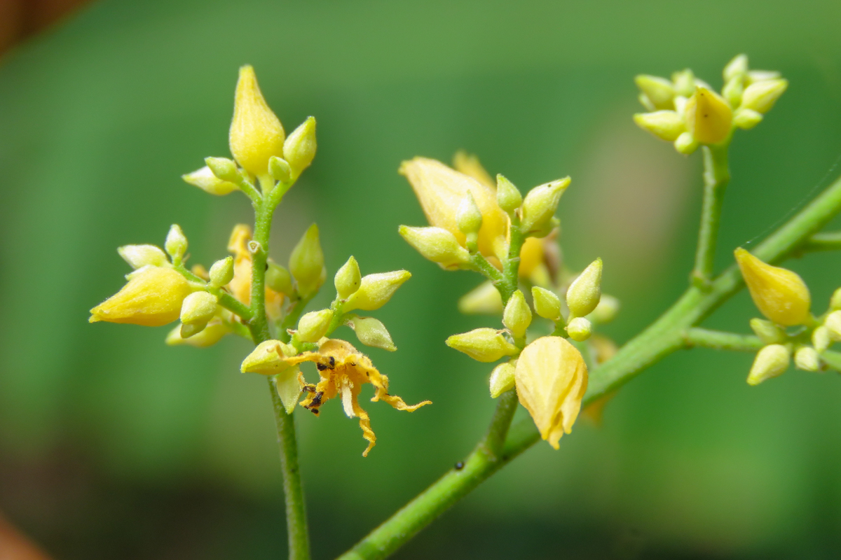
Fig. 1. H. brasiliensis’ flower (Bakewell-Stone, 2022).
Flowering of Hevea brasiliensis
Synchronous Flowering
Synchronous flowering is maintained over different rubber-growing regions and therefore cannot be linked to climate patterns, since these regions have different climates (Yeang, 2007a). In addition, flowering of H. brasiliensis near the equator is often bimodal, signifying the trees bloom twice a year. This further excludes the idea that flowering is linked to change in day length because when these processes are linked flowering only occurs once a year. As a result, the signal for synchronous flowering in H. brasiliensis could potentially be attributable to the following two environmental phenomena: bimodal advance in sunrise-sunset times measured against the chronometer and the bimodal variation in solar radiation intensity (Yeang, 2007a). The hypothesis proposing that synchronous flowering could be due to the sunrise-sunset advance measured against the chronometer suggests the photoreceptors of the plant can detect the time sunrise or sunset takes place. The plant is thought to be able to detect small time discrepancies which arise between solar time, based on the position of the sun and chronometer time, human clock time. Then, to use this difference timings to flower in sync in the most beneficial conditions. Due to plants being unaware of the precise chronometer time, this hypothesis is the less accepted one. At the equator, incoming solar radiation, also known as insolation, is entirely due to radiation intensity which is defined by the amount of solar energy that reaches a specific location. Hence, it is ultimately dependent on the angle of the sun since daylight is 12 hours all year round. Insolation at the equator peaks twice a year at the equinoxes, when the midday sun is directly above. H. brasiliensis trees flower when solar radiation intensity is high, probably detecting changes through photoreceptors like phytochromes, cryptochromes and phototropins. Due to H. brasiliensis having a low rate of ovaries developing into fruit after the successful pollination and fertilization, also known as fruit-set, the synchrony of flowering is important to increase the likelihood of effective pollination and ovary development (Yeang, 2007a). Thus, this can be seen as a design solution the tree has adapted to have to increase the rate of reproduction.
Plant Hormones and Nutrients Involved in Flowering
Gibberellin (GA) is a class of plant hormones that regulates critical processes like seed germination, stem elongation, leaf growth and most importantly, flowering. The biosynthesis of GA, seen in Figure 2, is a complex pathway which involves several catalytic enzymes that convert geranylgeranyl diphosphate (GGDP) into bioactive forms (Liu et al., 2025).

Fig. 2. Simplified GA biosynthetic pathway including the final stages of the 13-hydroxyl and non-13-hydroxyl pathways. The activity of several enzymes regulates the rate of GA biosynthesis, converting GA between different forms, of which only GA1, GA3, and GA4 exhibit bioactive properties (Pearce et al., 2013).
Nutrients also play an important role in plant physiology. They influence vegetative growth of H. brasiliensis trees as well as flower bud differentiation. The three essential nutrients needed in large amounts during plant growth and development are nitrogen, phosphorous and potassium. Trees in low-nutrient conditions generally exhibit delayed flowering compared to well-nourished ones. Low phosphorous levels and low gibberellin levels can postpone flowering (Liu et al., 2025). These pathways allow H. brasiliensis to regulate its nutrient use and hormonal balance to delay flowering and prioritize survival and growth under poor nutrient conditions.
Chemical Signaling and Maintenance of Homeostasis in H. brasiliensis
H. brasiliensis’ Adaptation to Different Light Intensity
The photosynthetic reaction remains the most important chemical process for H. brasiliensis, as it is responsible for supplying the main energy currency that all cells need. However, reactive oxygen species (ROS), which can cause oxidative stress and eventually damage cell integrity, are also byproducts of the light reaction and must be counteracted. Common ROS found in H. brasiliensis include superoxide (O₂⁻), hydrogen peroxide (H₂O₂), and the hydroxyl radical (OH-) (Wang, 2014). Most ROS in H. brasiliensis are formed due to electron leakage to O2, which is generated through splitting water in photosystem II. As shown in Figure 3, different electron transfers between reactants produce a variety of ROS.

Fig. 3. Different reaction pathways that produce different reactive oxygen species (Hajiboland, 2014).
Formation of O₂⁻ occurs in Photosystem I, where the electron-carrying ferredoxin has a steep negative electrochemical potential as the electron moves toward O₂ at the end of the electron transport chain. Subsequently, it oxidizes O₂ into the reactive O₂⁻. H₂O₂ is then formed through the dismutation reaction of O₂⁻, where the compound is simultaneously reduced and oxidized to produce H₂O₂ and O₂. Finally, the reaction between H₂O₂ and O₂⁻ generates the third most abundant ROS, the OH- (Hajiboland, 2014).
ROS are dangerous to cells because these highly reactive radicals can react with proteins, lipids, RNA, and DNA, disrupting cell integrity. These reactive species must be scavenged to maintain normal cellular function. One common method of scavenging involves using different target enzymes with different ROS. H. brasiliensis shows remarkable adaptability to varying light intensities and prevents oxidative stress under high light conditions. At high light intensity (1000 µmol/m²/s), the photosynthetic efficiency accelerates to its maximal rate, producing the greatest number of ROS as byproducts. Under this condition, light acts as a stimulus for the cisregulatory elements of antioxidant genes (Wang, 2014; Guo et al., 2025). This enhances the activity of enzymatic pathways by increasing the production of antioxidant enzymes, such as catalase and peroxidase. H. brasiliensis counters the increase of ROS due to higher photosynthetic rate with light-stimulated upregulation of antioxidant enzyme genes (Wang, 2014).
Apart from the enzymatic pathway, H. brasiliensis’ non-enzymatic pathways also neutralize reactive species. Typically, the most common non-enzymatic pathway is using carotenoid. The proximity between the triplet energy state of carotenoids and the singlet oxygen energy state allows for efficient electron exchange (Wang, 2014).
The β-carotene consists of a 40-carbon backbone with numerous conjugated double bonds; this allows for high reaction potential with the reactive oxygen species. As shown in Figure 4, each of these double bonds can react with and neutralize ROS. These double bonds interact with oxygen radicals, resulting in ground-state oxygen or H₂O₂ and excited carotenoids. Unlike reactive oxygen species, carotenoids can dissipate energy from their excited state through interactions with surrounding solvents without affecting cell integrity (Stahl & Sies, 2003).

Fig. 4. Neutralization of β-carotene with both singlet oxygen and any oxygen radicals. The plethora of double bond carbons give each carotene a lot of room to scavenge radicals (Stahl & Sies, 2003).
Here, H. brasiliensis’ maintain cellular integrity by preventing ROS accumulation from high frequency of photosynthesis through enzymatic and non-enzymatic pathways. These two mechanisms complement each other: although carotenoids are not produced in abundance like various antioxidant enzymes, it reacts quickly with reactive oxygen species, which provides sufficient time for enzyme synthesis and maximizes cellular protection.
Sugar Metabolism
Carbohydrates also play an important role in H. brasiliensis as a main product of the photosynthetic reaction. There are three main carbohydrates: sucrose, fructose, and glucose. Although polysaccharides are also present, starch and quebrachitol are detected only in small amounts. Among these carbohydrates, sucrose is the most abundant in mature leaves, with a sucrose-to-starch ratio of approximately 11, the highest reported among plants to date. This value can be compared to tapioca plant, another member of the Euphorbiaceae family, which has a sucrose-to-starch ratio of about 5 (Zhu et al., 2018).
To maintain this high sucrose concentration in its leaves, H. brasiliensis relies on a complex sucrose-cleaving and sucrose-synthesizing enzymatic system. In total, there are three sucrose-synthesizing enzymes and four sucrose-cleaving enzymes. Sucrose-synthesizing enzymes are primarily active in mature leaves. These include sucrose-phosphate synthase (SPS), which converts UDP-glucose and fructose-6-phosphate into sucrose-6-phosphate; sucrose-phosphate phosphatase (SPP), which removes the phosphate group; and sucrose synthase acting in the synthesis direction (SuSy), which produces sucrose in a transportable form.
Sucrose-cleaving enzymes are mainly detected in young leaves and include four types: cell wall invertase (CWI), vacuolar invertase (VIN), cytoplasmic invertase (NIN), and sucrose synthase acting in the cleavage direction (SuSy) (Zhu et al., 2018).
H. brasiliensis’ leaves are categorized into four stages, with stage I, II, and III (bronze and light green) being viewed as immature leaves, and IV being viewed as mature leaves (dark green). This categorization makes the different sucrose enzyme roles in young and mature leaves more apparent. Of the seven enzymes identified, all sucrose-synthesizing enzymes and one sucrose-cleaving enzyme (SuSy, acting in both directions; SPS; SPP) are found in the stage IV mature leaves of H. brasiliensis. The requirement for such a high concentration of sucrose arises from the mature leaves’ role as source tissues, as they export sucrose to young leaves and laticifers system via the phloem. Meanwhile, the metabolic demands of young leaves explain why they contain only sucrose-cleaving enzymes (CWI, VIN, NIN, SuSy). CWI allows sucrose importation into the young leaves, while VIN hydrolyzes sucrose within the vacuole, breaking it into fructose and glucose in the vacuole, creating a gradient that leads to osmotic water uptake, increasing the turgor pressure to drive cellular expansion in young leaves (Sergeeva et al., 2006; Zhu et al., 2018). NIN, located in the cytosol, functions as a signaling enzyme that regulates sugar homeostasis through feedback inhibition of sucrose. SuSy, acting in the cleavage direction breaks down sucrose into UDP-glucose and fructose, supplying substrates for cell wall synthesis and glycolysis (Zhu et al., 2018). This enzyme distribution explains the high sucrose-to-starch ratio of 11 found in mature leaves. As shown in Figure 5, the mature leaves contain the highest sucrose concentration, emphasizing their role as the tree’s primary source tissue.

Fig. 5. Quantity of four kinds of carbohydrate present in each stage of the leaf. Sucrose content is the highest among stage IV leaves (Zhu et al., 2018).
H. brasiliensis leaves have a conflicting demand between growth and defense. Immature leaves rely on hydrolysis of sucrose into fructose and glucose for cell expansion while there is a concurrent demand to supply sucrose to the laticifer network for protection against microbes and herbivores. To satisfy both needs, H. brasiliensis develops a unique compartmentalization of mainly sucrose-cleaving enzymes in young leaves and sucrose-synthesizing enzymes in mature leaves. This metabolic partitioning ensures a steady supply of sucrose to the laticifer network while also sustaining the growth of developing foliage.
The Symbiotic Relation of Hevea Brasiliensis with Arbuscular Mycorrhizal Fungi
Located in tropical environments, H. brasiliensis is exposed to recurrent dry seasons, and periods of nutrient and water drought. Sharing the territory with other species, competition for limited resources in the soil during these harsh periods is inevitable. To get its share of nutrients, the rubber tree has developed a symbiotic relationship with plant-growth favoring fungi in the soil.
Voluntary Colonization by Fungi
Arbuscular Mycorrhizal Fungi (AMF), as seen in Figure 6, establish a symbiotic relationship with a plant host, where they receive carbohydrates produced from photosynthesis in exchange for access to their hyphal network. Hyphae are branched, tubular filaments that extend from the fungus to absorb nutrients and water.

Fig. 6. The root network of arbuscular mycorrhizal fungi (AMF), with rhizosphere showing soil surrounding the root (influenced by root metabolism) and hyphosphere hosting network that extends to absorb nutrients from soil nearby and interact with other organisms (Faghihinia et al., 2022).
Though there is a great variety of AMF, H. brasiliensis seems to have developed a consistent relationship with three main fungi: Glomus, Aculospora, and Entrophospora (Gao et al., 2021). Table 1 shows the frequency of different AMF species found in the rhizosphere of H. brasiliensis.
Table 1. Frequency of AMF genera/species in the rhizosphere of H. brasiliensis at 11 sampling sites on Hainan Island, adapted from (Gao et al., 2021).
| Genus | Number of species | Sites detected | Frequency (%) |
|---|---|---|---|
| Archaeospora | 1 | 2 | 18.2 |
| Glomus | 43 | 11 | 100 |
| Acaulospora | 18 | 11 | 100 |
| Entrophospora | 3 | 4 | 36.4 |
| Gigaspora | 1 | 1 | 9.1 |
| Scutellospora | 2 | 2 | 18.2 |
H. brasiliensis’ roots, when in water or nutrient deficiency, exude strigolactones (Figure 7), which attracts AMF spores and stimulates the growth of the fungus towards the root (Farhaoui et al., 2025). As it approaches, the AMF releases in turn CO4 or CO5 lipo-chitooligosaccharides “Myc-signals” into the soil. Because these resemble pathogen-associated molecular patterns closely, they will both trigger a symbiotic and slightly defensive response from the roots once H. brasiliensis’ receptors receive the “Myc-signals” (Farhaoui et al., 2025).

Fig. 7. Illustration of basic strigolactone structure, (adapted from National Center for Biotechnology Information, 2025).
The symbiotic response occurs as the fixation of the fungus’ hyphae onto Hevea’s roots’ epidermous cells and their differentiation into appressoria (Figure 8). These act as entry points for the fungus’ hyphae, that penetrate the cortex with wall-breaching enzymes such as laccases, chitinases or glucanases, and mechanical stress. Once inside the cortex, the hyphae branch into symbiotic interfaces: the arbuscules, and the vesicles. Arbuscules work as nutrient exchange platforms where nutrients like phosphorus (P) are given to the H. brasiliensis in return for carbon metabolites from photosynthesis (Farhaoui et al., 2025).

Fig. 8. Schematic illustration of the symbiotic system of arbuscular mycorrhizal fungi (AMF) in the host plant root (Farhaoui et al., 2025).
The defensive response occurs as the formation of a peri-arbuscular membrane, that prevents further incursion by the fungus. Mechanically, this process is very similar to other defensive processes H. brasiliensis employs against pathogenic fungi.
In an experiment by Nicole et al., (Nicole et al., 1991) fluorescence highlights the activity of cinnamyl alcohol dehydrogenase (CAD) and isoperoxidase (PO), both enzymes that participate in H.brasiliensis’ cell wall reinforcement, like lignin production and phellogen (meristematic cell layer responsible for development of periderm) activity. When exposed to cell-wall extracts of root rot fungus Rigidoporus lignosus, CAD activity and lignin synthesis greatly increased within four days of inoculation and remained high for five weeks before reconverging to control values (Figure 9). This highlights both the highly efficient, and long-lasting reaction mechanism H. brasiliensis has against invasive fungi species. In the rhizosphere of the roots, there are naturally a plethora of pathogenic fungi, and H. brasiliensis is capable of quickly recognizing these signals, then set up a strengthened cell wall against it for five weeks.

Fig. 9. Cinnamyl-alcohol dehydrogenase activity (CAD) in roots after treatment with elicitors (ELI 1, 2, 3, 4). □, control 1, healthy plants; ○, control 2, plants wounded with a needle; ∆, control 3, plants injected with water; X , ELI 1 (initial elicitor), black □ ELI 2 (pronase-treated); black ○, ELI 3 (ethanol-soluble); black ∆, ELI 4 (pronase-treated and ethanol soluble)(Nicole et al., 1991).
Table 2: Stimulation of phellogen activity in roots of H. brasiliensis at different times following treatments with Rigidoporus lignosus elicitors (ELI I, 2, 3, and 4) (Nicole et al., 1991).
Days after elicitor treatment | ||||||
|---|---|---|---|---|---|---|
| Assay | 3 | 5 | 8 | 15 | 30 | 45 |
| ELI 1 | 0 | 0 | 0 | 6 | 2 | 2 |
| ELI 2 | 2 | 0 | 1 | 1 | 2 | 0 |
| ELI 3 | 0 | 0 | 2 | 11 | 0 | 0 |
| ELI 4 | 2 | 0 | 0 | 0 | 2 | 0 |
| C2 | 0 | 1 | 2 | 0 | 0 | 0 |
| C3 | 0 | 1 | 0 | 0 | 0 | 1 |
| nC1 | 4-5 | 5-6 | 7-9 | 9-11 | 11-13 | 13-16 |
In comparison, a similar experiment by Schwob et al., using the common H. brasiliensis AMF Glomus mosseae, found very close results to the previous study. Cinnamyl alcohol dehydrogenase (CAD) and isoperoxidase activity peaked at two weeks after inoculation, when the fungus first reached the cortex, before going back to control values (Figure 10). However, it was observed that scopoletin quantity, a phytoalexin induced by pathogenic fungi, did not vary between control and inoculated subjects. Hence, we can see how H. brasiliensis, through different chemical reactions, uses the same defense system differently for friendly and pathogenic fungi (Schwob et al., 2000).

Fig. 10. Enzyme activities, and lignin and scopoletin contents in inoculated and control root tissues of rubber trees during the first five weeks after inoculation. A: CAD activity; B: PO activity; C: lignin percentage; D: scopoletin percentage. Means with the same letter are not different by Scheffe's test (p<0.05) (Schwob et al., 2000).
H. brasiliensis has the same early-stage response to both friendly and pathogenic species. It automatically treats the cell wall breach of both kinds of fungi as a potential attack, causing CAD and peroxidase to strengthen the cell walls and prevent damage. The immediate response of the tree prioritizes survival and prevents infection, giving time for the tree to recognize the type of fungus involved. In the case of root-rotting fungi, a long-lasting cell wall reinforcement is maintained. Meanwhile, the cell wall reinforcement declines, and permeability increases to allow nutrient exchange in the presence of arbuscular mycorrhizal fungi. This system allows both optimal protection of the tree and minimizes energy expenditure by using the same defense mechanism but triggering different follow-up chemical reactions.
Role of AMF During Drought or Lack of Nutrients
In nature, H. brasiliensis roots mostly absorb soil-solution inorganic compounds containing P, which represents around 1% of the total P in the soil, which are either absorbed onto the soil surfaces or precipitated as Fe and Al phosphates in acid soils and as Ca and Mg phosphates in more alkaline and calcareous soils. Most of this small reserve of P is usually absorbed through diffusion of P in the soil following a concentration gradient, but only the P close to the root is taken by the tree, attributable to the slow diffusion rate of P in the soil, which is about 10 -11 to 10 -8 cm²/s, compared to the fast diffusion rate of 0.89 x 10 -5 cm2/s of water (Bolan, 1991).
However, H. brasiliensis’ symbiotic bond with AMF allows an increase in the inflow of P into the tree, from 3.6×10⁻¹⁴ mol⋅cm-1⋅s-1 to 17.0×10⁻¹⁴ mol⋅cm-1⋅s-1. There are multiple possible reasons for this. Firstly, AMF’s hyphae extend the possible exploration of the soil with their entire network, also extending the diffusion zone (area where P is absorbed) around 60-fold. Secondly, since hyphae hairs are much finer than root hairs, once inaccessible cracks in soil compactions become tappable for P. Finally, it is hypothesized that arbuscular mycorrhizal fungi have higher affinity for P absorption and a lower concentration threshold, allowing H. brasiliensis to absorb less concentrated sources of P in the soil (Bolan, 1991).
AMFs are also useful to the rubber tree in periods of water drought during dry seasons. Through mutualistic interaction with the fungi, H. brasiliensis can utilize AMF’s hyphal network that allows larger soil exploration and increased absorption of water. As seen in Figure 11, AMF enhances characteristics of H. brasiliensis in a well-watered (WW) state and decreases the impact on plant growth in a water deficient (WD) state. The photosynthetic capabilities are maintained throughout water deficiency. Chlorophyll a, chlorophyll b, and total chlorophyll concentrations in the leaf tissues of AMF-inoculated plants under WD declined by 33.6%, 23.0%, and 27.9% of WW, respectively. Meanwhile, in non-inoculated plants, a sharp decline by 51.6%, 36.4%, and 43.3% over the control was observed over 14 days. Moreover, the photosynthetic rate of AMF inoculated plants only dropped by 5.7% in that period, whereas non-inoculated plants’ photosynthetic rate dropped by 7.7% (Tisarum et al., 2022).

Fig. 11. Growth parameters of Hevea brasiliensis inoculated with (+) or without (-) arbuscular mycorrhizal fungi (AMF), , and subsequently subjected to well-watered (WW) and water deficit (WD) conditions for 14 days, adapted from (Tisarum et al., 2022).
Essentially, H. brasiliensis resolves the issue of lack of P it can absorb due to the naturally slow diffusion rate of phosphate in the soil with interactions with arbuscular mycorrhizal fungi. Instead of developing new root structures, or specialized organ to uptake phosphate efficiently, and be more drought-resistant, it takes advantage of its pre-existing defense system against pathogenic fungi, modifying it slightly to instead allow nutrient exchange, and integrate the arbuscular mycorrhizal fungi’s extensive hyphal network to its advantage.
Latex Coagulation
Overview of the Mechanism of Hevea Latex Coagulation
When latex exudes out of the rubber tree, a series of biochemical reactions that destabilize and aggregate the rubber particles are triggered. This changes both the components and colloidal stability of latex which includes mechanical stability, sensitivity of the latex to mechanical agitations, and chemical stability, sensitivity to addition of ionic material (Bowler, 1953; de Vries, 1923; Ng et al., 2022). Cessation of latex flow and plugging of the wound with coagulation prevents metabolite loss and is vital for plant defense against pathogen invasion (Gidrol et al., 1994; Wititsuwannakul et al., 2008).
First Stage: Destabilization of Rubber Particles
Hevea latex is a charged colloidal suspension of rubber particles dispersed in a cytoplasmic aqueous medium of nonrubber components, which include proteins, lipids, carbohydrates, and inorganic salts, also known as C-serum (Kumarn et al., 2018; Ng et al., 2022). In each rubber particle, there are several hundred rubber molecules, cis-1,4-polysoprene molecules, with a wide range of molecular size distribution. The α-end of the rubber molecule is capped with a mono- (OP) or diphosphate (OPP) group, while the ω-end is an isoprene-derived group next to two trans-1,4-isoprene units connecting the main polyisoprene chain (Figure 12) (Kumarn et al., 2018; Wititsuwaannakul et al., 2004).

Fig. 12. Two different structures based on the α-end of the rubber molecule (Kumarn et al., 2018).
During biosynthesis, the rubber chain is elongated inward, pointing the ω-end outward. As for the α-end capped with a hydrophilic group, it is linked with a phospholipid and placed to face the C-serum, minimizing polar-nonpolar interaction (Figure 13) (Kumarn et al., 2018). The colloidal stability of the system of rubber particles is attributable to their negatively charged protein-phospholipid membrane layer which creates a repulsive environment that prevents collision between particles (Ng et al., 2022; Wititsuwaannakul et al., 2004). While anionic proteins located in the C-serum provide an electrostatic effect to the medium, stabilization is further enhanced with the absorption of polar lipids and free fatty acids to the rubber particle membrane. These negatively charged species adhere to the membrane by anchoring their hydrophobic tails inward and exposing their charged heads to the C-serum (Figure 13) (Kumarn et al., 2018; Ng et al., 2022).

Fig. 13. Model of the Hevea rubber particle in latex (Kumarn et al., 2018).
As rubber particles, lutoids are protected by a negatively charged protein-phospholipid membrane. At the equilibrium of electrostatic repulsion, lutoids and rubber particles do not collide in C-serum (Figure 14a). However, the second highly acidic colloidal system inside lutoids, B-serum, is responsible for the destabilization of the negatively charged colloidal suspension of rubber particles. As put forward in Essay 1, change of osmotic gradient due to biochemical actions, bacteria action, or tapping can puncture lutoids that are osmotically sensitive (Priyadarshan, 2017). The breakage of the lutoids is followed by the release of B-serum and its destabilizing attributes, which include divalent cations, like Mg2+ and Ca2+, acidic pH, positively charged proteins, and acid hydrolases (Figure 14b). The destabilizing attributes neutralize the surface of lutoids and rubber particles (Ng et al., 2022; Yusuf et al., 2024). Once the isoelectric point, the pH at which the negatively and positively charged species are present in equal concentration of latex, is achieved, the two colloidal systems are disturbed and come into contact (LibreTexts, n.d.) (Figure 14c). Collision breaks their membrane layers and allows the adhesion between particles, a process known as flocculation (Ng et al., 2022).

Fig. 14. The process of latex destabilization leading to flocculation. (a) Stabilization of the colloidal suspension of rubber particles and lutoids due to repulsive forces. (b) Release of B-serum following the breakage of the lutoid membrane. (c) Collision between rubber particles and lutoids (Ng et al., 2022).
Second Stage: Aggregation of Rubber Particles
Three different models have been put forward to explain the mechanism of rubber particle aggregation: coagulation via binding of hevein, coagulation by Hevea lectin-like protein (HLL), and coagulation by the combinatorial effects of chitinase and glucanase (Ng et al., 2022).
According to the first model of 1994, the formation of latex coagulum is due to multivalent bridges between hevein and rubber particles (Gidrol et al., 1994). Representing more than 20 % of total soluble latex proteins, hevein is a 43 amino acid residue polypeptide comprising of two strands of β-sheet and three α-helices stabilized by four disulfide bridges (Figure 15A) (Berthelot et al., 2016). Hevein is a merolectin, which is a protein with a single carbohydrate-binding domain (Figure 15B) (Van Damme et al., 2007).

Fig. 15. Structure of hevein. (A) Structure of hevein where N and C indicate the N-terminal and C-terminal ends of the polypeptide chain (B) Binding of poly- or monosaccharides to the carbohydrate-binding domain. H-bonds anchoring the oxygens of the sugars to the amino acid residues of the binding domain are represented by the dashed lines. In orange, aromatic residues involved in stacking interaction with the sugars are highlighted (Van Damme et al., 2007).
Figure 16A demonstrates the mechanism behind aggregation, which consists of the creation of multivalent bridges between rubber particles by hevein through its binding to the N-acetyl-D-glucosamine, the monomeric unit of chitin, moiety of the 22kDa receptor glycoprotein located on the surface of rubber particles. The binding affinity of hevein is regulated by the concentration of Ca2+ and the enzyme chitinase. It was determined that hevein could bind to the N-acetyl-D-glucosamine moiety only in the presence of Ca2+ physiological concentrations of 200µM. By binding to hevein, Ca2+ favors the dimerization of hevein, allowing it to bind to two saccharides, and stabilizes the protein against the hydrolytic activity of proteolytic enzymes in B-serum. Hevein creates multivalent bridges between rubber particles when concentrations of enzymes chitinase or N-acetyl-β-D-glucosaminidase are low. High chitinase activity inhibits coagulation by removing the N-acetyl-D-glucosamine moiety from the 22kDa receptor protein, which increases the concentration of free N-acetyl-D-glucosamine and can saturate the binding sites of hevein still present in the cytosol (Figure 16A) (Gidrol et al., 1994).

Fig. 16. Representation of the model of coagulation via binding of hevein. (A) Low chitinase and N-acetyl-β-D-glucosaminidase activity. (B) High chitinase and N-acetyl-β-D-glucosaminidase activity (Gidrol et al., 1994).
In 2008, a new model was put forward. According to this model, the bursting of lutoids releases the intrinsic latex lectin (HLL) located on the lutoid membrane (Figure 17). HLL can then bind to either the rubber particle HLL binding protein (RP-HLLBP), also known as Hev b 3, or the C-serum HLL binding protein (CS-HLLBP), also referred to as Hev b 7. RP-HLLBP bound to the rubber particle surface acts as the ligand for HLL to cause aggregation (Figure 17a). CS-HLLBP acts as an anti-coagulating factor that competes for HLL, which prevents aggregation and maintains the colloidal stability of the latex (Figure 17b). These two proteins therefore have a dynamic competitive interaction with HLL. This model states that the formation of latex coagulum is mainly dependent on the interactions between HLL and RP-HLLBP and not those between HLL and CS-HLLBP. Therefore, for coagulum to form, the number of exposed HLL binding sites has to exceed those to be occupied by CS-HLLBP (Figure 17a) (Wititsuwannakul et al., 2008).

Fig. 17. Representation of the model of coagulation by HLL. (a) Number of interactions between HLL and RP-HLLBP exceed those between HLL and CS-HLLBP. (b) Number of interactions between HLL and CS-HLLBP exceed those between HLL and RP-HLLBP. SRP, small rubber particle (Wititsuwannakul et al., 2008).
The most recent proposed model includes all the main proteins involved in lutoid-mediated rubber particle aggregation (RPA). Figure 18C shows that glucanase, a vascular glycoprotein, enhances RPA. As stated in the first model, inhibition of RPA by chitinase and enhancement of RPA by hevein is showcased in figure 18D-E. The addition of chitinase inhibited RPA induced by hevein and strongly enhanced RPA induced by glucanase (Figure 18F-G). On the other hand, the addition of hevein to glucanase had little effect (Figure 18H). All of this suggests that these three proteins act as positive activators of RPA. Where the first model stated that chitinase acts as an inhibitor of RPA, this model states that chitinase indirectly enhances RPA with its interaction with glucanase (Wang et al., 2013).

Fig. 18. Results of RPA induced by several lutoid proteins in increasing amounts from lanes 1-6. (A) Primary lutoid solution (PLS). (B) Secondary lutoid protein solution (SLS). (C) Purified glucanase. (D) Hevein. (E) Chitinase. (F) 50 µg of hevein with 0-75 µg of chitinase. (G) 50 µg of glucanase with 0-75 µg of chitinase. (H) 50 µg of glucanase with 0-75 µg of hevein. Primary lutoids are found in primary laticifers, also known as articulated laticifers. Secondary lutoids are found in secondary laticifers or nonarticulated laticifers. To establish relative RPA, the lutoid proteins were added to rubber particle suspensions and stained with basic fuchsin. A-H constitute the representative results and a-h are the corresponding statistical results (Wang et al., 2013).
Based on the results presented above and in-depth proteomics of lutoids, a possible revised model for lutoid-mediated RPA and latex coagulation was presented in Figure 19. Upon wounding, latex flows out of laticifers, resulting in a turgor pressure decrease leading to bursting of lutoids. Then, lutoid inclusion-localized hevamine, a defense protein like chitinase, contacts membrane-localized glucanase, facilitating RPA, and the released chitinase interacts with glucanase to induce glucanase-mediated RPA. According to this model, hevein, prohevein, and lectin-like protein are still part of RPA. However, it is important to note that this model differs from the HLL coagulation model regarding the nature of the latex lectin-like protein. The newer model states that HLL could not be a key activator for RPA because, although lectin is present in numerous plants, fungi, bacteria, and algae, it is not relatively abundant in latex lutoids. Moreover, lectins from non-latex-producing plants and animals have shown to be ineffective at inducing RPA. Finally, it is difficult to explain the results of Figure 19A-B, where protein solutions from primary and secondary lutoids induced RPA to a high degree even with a low abundance of lectin, if lectin is really the common key activator of RPA. Considering all this, the authors suggested that latex lectin-like protein may not be actual latex lectin, but prohevein, which is abundant in lutoids (Wang et al., 2013). Since HLL, 17kDa, has a similar size to prohevein, 18.5kDa, and chitin affinity, it is possible that HLL could be in fact prohevein. Hevein is formed from the larger protein prohevein, a 187 amino acid protein cleaved into two fragments, which include the N-terminal 43 amino acid hevein and a C-terminal domain (Berthelot et al., 2016). As lectins, both hevein and prohevein display various agglutination properties, have high chitin-binding activity, and interact selectively and noncovalently with chitin. Therefore, according to the newest model and the results of Figure 18D, hevein and prohevein are the lectin-like proteins, which, when released from lutoids, can interact with a glycoprotein on the rubber particle surface leading to coagulation. As demonstrated in Figure 18F, this effect can still be inhibited by chitinase, which acts as an inhibitor by releasing N-acetyl-D-glucosamine moieties to block the hevein-binding site of hevein and in the N-terminal of prohevein (Berthelot et al., 2016; Wang et al., 2013).

Fig. 19. Illustration of the localization and functions of the main lutoid proteins with the proposed model of lutoid-mediated RPA leading to latex coagulation. The red arrows pointing toward RPA depart from glucanase and chitinase, the two new proteins identified to be involved in RPA according to this model. The black arrows between glucanase and hevamine represent their interaction, enhancing glucanase-mediated RPA. The black arrows linking lectin and prohevein represent the mechanism of coagulation via RPA in a lectin-like manner, still valid according to the model. LRP, large rubber particle; PLC, primary laticifer cell; ILP, inclusions of lutoids from primary laticifers; MLP, membranes of lutoids from primary laticifers; MLS, membranes of lutoids from secondary laticifers; ILS, inclusions of lutoids from secondary laticifers; SLC, secondary laticifer cell; SRP, small rubber particle (Wang et al., 2013).
Conclusion
Even though H. brasiliensis is located near the equator, where there is a lack of distinct meteorological signs, it’s known to have a low rate of fruit-set, and it can synchronize their flowering to maximize pollination. Through photoreceptors, they detect biannual peaks of insolation, which the trees use as signals for synchronous flowering. In tropical climates, where dry seasons and periods of drought are common occurrences, H. brasiliensis has established a symbiotic relationship with arbuscular mycorrhizal fungi to increase the inflow of phosphorus and water in periods of drought. Although photosynthesis supplies energy to H. brasiliensis, by-products cause oxidative stress, which damages cell integrity. While high light intensity maximizes the yield of the ROS, it also enhances the activity of enzymatic pathways that scavenge ROS. Other non-enzymatic pathways involving highly reactive species that neutralize ROS are also present to allow a faster response and provide sufficient time for antioxidant enzymes synthesis. In its mature leaves, H. brasiliensis has the highest sucrose-to-starch ratio. Such a high concentration of sucrose in mature leaves allows them to export sucrose to young developing leaves and to laticifers, where latex biosynthesis occurs. This distinctive concentration is due to a compartmentalization pattern, in which sucrose-cleaving enzymes are primarily located in young leaves, and sucrose-synthesizing enzymes are concentrated in mature leaves. Finally, it is known that its high latex yield sparked the rubber industry’s interest in the H. brasiliensis. Latex, when not exuding, remains stabilized due to a repulsive environment created by the negatively charged membranes of rubber particles and lutoids. Once injured, latex flows out of the rubber tree and plays a defensive role through, among many other ways, wound plugging. More than a barrier to possible pathogen invasion, wound plugging is important to the tree to prevent faster stoppage of metabolite loss than what only a decrease in turgor pressure would allow. Proteins are involved in this faster wound plugging through destabilization of rubber particles and formation of coagulum. Altogether, these design solutions showcase how the rubber tree has mastered its ability to optimize nutrients intake and distribution to meet the metabolic demands of the reproductive and defensive systems.
References
References
Bakewell-Stone, P. (2022). Hevea brasiliensis (rubber). CABI Compendium. https://doi.org/10.1079/cabicompendium.27999
Berthelot, K., Peruch, F., & Lecomte, S. (2016). Highlights on Hevea brasiliensis (pro)hevein proteins. Biochimie, 127, 258-270. https://doi.org/https://doi.org/10.1016/j.biochi.2016.06.006
Bolan, N. (1991). A critical review on the role of mycorrhizal fungi in the uptake of phosphorus by plants. Plant and Soil, 134(2), 189-207.
Bowler, W. W. (1953). Electrophoretic Mobility Study of Fresh Hevea Latex. Industrial & Engineering Chemistry, 45(8), 1790-1794. https://doi.org/10.1021/ie50524a050
Britannica Editors (2025, September 19). lignin. Encyclopedia Britannica. https://www.britannica.com/science/lignin
de Vries, O. (1923). Coagulation phenomena in hevea latex. Recueil des Travaux Chimiques des Pays-Bas, 42(8), 701-705. https://doi.org/https://doi.org/10.1002/recl.19230420811
Faghihinia, M., Jansa, J., Halverson, L. J., & Staddon, P. L. (2022). Hyphosphere microbiome of arbuscular mycorrhizal fungi: A realm of unknowns. Biology and Fertility of Soils, 59(1), 17–34. https://doi.org/10.1007/s00374-022-01683-4
Farhaoui, A., Taoussi, M., Laasli, S.-E., Legrifi, I., El Mazouni, N., Meddich, A., Hijri, M., & Lahlali, R. (2025). Arbuscular Mycorrhizal Fungi and Their Role in Plant Disease Control: A State-of-the-Art. The Microbe, 100438. https://doi.org/10.1016/j.microb.2025.100438
Gao, X., Lv, J., Guo, C., Hu, A., Wu, X., & Li, Z. (2021). Species diversity of arbuscular mycorrhizal fungi in the rhizosphere of Hevea brasiliensis in Hainan Island, China. Phyton, 90(1), 179. https://doi.org/10.32604/phyton.2021.012968
Gidrol, X., Chrestin, H., Tan, H. L., & Kush, A. (1994). Hevein, a lectin-like protein from Hevea brasiliensis (rubber tree) is involved in the coagulation of latex. Journal of Biological Chemistry, 269(12), 9278-9283. https://doi.org/https://doi.org/10.1016/S0021-9258(17)37104-1
Guo, B., Fan, S., Liu, M., Yang, H., Dai, L., & Wang, L. (2025). ATP synthase members of chloroplasts and mitochondria in rubber trees (Hevea brasiliensis) response to plant hormones. Plants, 14(4), 604. https://doi.org/10.3390/plants14040604
Hajiboland, R. (2014). Reactive oxygen species and photosynthesis. Oxidative Damage to Plants, 1–63. https://doi.org/10.1016/b978-0-12-799963-0.00001-0
Kumarn, S., Churinthorn, N., Nimpaiboon, A., Sriring, M., Ho, C.-C., Takahara, A., & Sakdapipanich, J. (2018). Investigating the Mechanistic and Structural Role of Lipid Hydrolysis in the Stabilization of Ammonia-Preserved Hevea Rubber Latex. Langmuir, 34(43), 12730-12738. https://doi.org/10.1021/acs.langmuir.8b02321
LibreTexts. (n.d.). 25.3: Isoelectric Points and Electrophoresis. Chemistry LibreTexts. https://chem.libretexts.org/Bookshelves/Organic_Chemistry/Map%3A_Organic_Chemistry_(Wade)_Complete_and_Semesters_I_and_II/Map%3A_Organic_Chemistry_(Wade)/25%3A_Amino_Acids_Peptides_and_Proteins/25.03%3A_Isoelectric_Points_and_Electrophoresis#title
Liu, M., Yang, H., Fan, S., Fan, X., Guo, B., Dai, L., Lin, Q., & Wang, L. (2025). Transcriptomic analysis reveals the gibberellin signaling pathway involved in the regulation of flower number in Hevea brasiliensis Muell. Arg. Industrial Crops and Products, 235, 121697. https://doi.org/10.1016/j.indcrop.2025.121697
National Center for Biotechnology Information (2025). PubChem Compound Summary for CID 46174042, Strigolactone ABC-rings. Retrieved October 14, 2025 from https://pubchem.ncbi.nlm.nih.gov/compound/Strigolactone-ABC-rings.
Ng, J. W., Othman, N., & Yusof, N. H. (2022). Various coagulation techniques and their impacts towards the properties of natural rubber latex from Hevea brasiliensis — a comprehensive review related to tyre application. Industrial Crops and Products, 181, 114835. https://doi.org/https://doi.org/10.1016/j.indcrop.2022.114835
Nicole, M., Toppan, A., Geiger, J.-P., Roby, D., Nandris, D., & Rio, B. (1991). Defense responses of Hevea brasiliensis to elicitors from root rot fungi. Canadian journal of botany, 69(8), 1819-1824.https://doi.org/10.1139/b91-231
Pearce, S., Vanzeg, L., & Dubcovsky, J. (2013). Exogenous gibberellins induce wheat spike development under short days only in the presence of VERNALIZATION1. Plant physiology, 163. https://doi.org/10.1104/pp.113.225854
Priyadarshan, P. M. (2017). Latex Production, Diagnosis and Harvest. In P. M. Priyadarshan (Ed.), Biology of Hevea Rubber (pp. 51-82). Springer International Publishing. https://doi.org/10.1007/978-3-319-54506-6_5
Schwob, I., Ducher, M., & Coudret, A. (2000). An arbuscular mycorrhizal fungus (Glomus mosseae) induces a defence-like response in rubber tree (Hevea brasiliensis) roots. Journal of plant physiology, 156(2), 284-287. https://doi.org/10.1016/S0176-1617(00)80319-3
Sergeeva, L. I., Keurentjes, J. J., Bentsink, L., Vonk, J., van der Plas, L. H., Koornneef, M., & Vreugdenhil, D. (2006). Vacuolar invertase regulates elongation of Arabidopsis thaliana roots as revealed by QTL and Mutant Analysis. Proceedings of the National Academy of Sciences, 103(8), 2994–2999. https://doi.org/10.1073/pnas.0511015103
Stahl, W., & Sies, H. (2003). Antioxidant activity of carotenoids. Molecular Aspects of Medicine, 24(6), 345–351. https://doi.org/10.1016/s0098-2997(03)00030-x
Tisarum, R., Samphumphuang, T., Yooyoungwech, S., Singh, H. P., & Cha-Um, S. (2022). Arbuscular mycorrhizal fungi modulate physiological and morphological adaptations in para rubber tree (Hevea brasiliensis) under water deficit stress. Biologia, 77(7), 1723-1736. https://doi.org/10.1007/s11756-022-01016-8
Van Damme, E. J. M., Rougé, P., & Peumans, W. J. (2007). 3.26 - Plant Lectins. In H. Kamerling (Ed.), Comprehensive Glycoscience (pp. 563-599). Elsevier. https://doi.org/https://doi.org/10.1016/B978-044451967-2/00067-2
Wang, L. (2014). Physiological and molecular responses to variation of light intensity in rubber tree (Hevea brasiliensis Muell. arg.). PLoS ONE, 9(2). https://doi.org/10.1371/journal.pone.0089514
Wang, X., Shi, M., Wang, D., Chen, Y., Cai, F., Zhang, S., Wang, L., Tong, Z., & Tian, W.-M. (2013). Comparative Proteomics of Primary and Secondary Lutoids Reveals that Chitinase and Glucanase Play a Crucial Combined Role in Rubber Particle Aggregation in Hevea brasiliensis. Journal of Proteome Research, 12(11), 5146-5159. https://doi.org/10.1021/pr400378c
Wititsuwaannakul, D., Rattanapittayaporn, A., Koyama, T., & Wititsuwaannakul, R. (2004). Involvement of Hevea Latex Organelle Membrane Proteins in the Rubber Biosynthesis Activity and Regulatory Function. Macromolecular Bioscience, 4(3), 314-323. https://doi.org/https://doi.org/10.1002/mabi.200300080
Wititsuwannakul, R., Pasitkul, P., Jewtragoon, P., & Wititsuwannakul, D. (2008). Hevea latex lectin binding protein in C-serum as an anti-latex coagulating factor and its role in a proposed new model for latex coagulation. Phytochemistry, 69(3), 656-662. https://doi.org/https://doi.org/10.1016/j.phytochem.2007.09.021
Wititsuwannakul, R., Pasitkul, P., Kanokwiroon, K., & Wititsuwannakul, D. (2008). A role for a Hevea latex lectin-like protein in mediating rubber particle aggregation and latex coagulation. Phytochemistry, 69(2), 339-347. https://doi.org/https://doi.org/10.1016/j.phytochem.2007.08.019
Yeang, H.-Y. (2007a). The sunshine-mediated trigger of synchronous flowering in the tropics: the rubber tree as a study model. New Phytologist, 176(4), 730-735. https://doi.org/10.1111/j.1469-8137.2007.02258.x
Yeang, H.-Y. (2007b). Synchronous Flowering of the Rubber Tree (Hevea brasiliensis) Induced by High Solar Radiabon Intensity. The New Phytologist, 175(2), 283-289. https://www.jstor.org/stable/4641046
Yusuf, M., Ridha, S., & Kamyab, H. (2024). Recent progress in NP-Based Enhanced oil Recovery: Insights from molecular studies. Journal of Molecular Liquids, 396, 124104. https://doi.org/https://doi.org/10.1016/j.molliq.2024.124104
Zhu, J., Qi, J., Fang, Y., Xiao, X., Li, J., Lan, J., & Tang, C. (2018). Characterization of sugar contents and sucrose metabolizing enzymes in developing leaves of Hevea brasiliensis. Frontiers in Plant Science, 9. https://doi.org/10.3389/fpls.2018.00058