
Table of Contents
Keywords: eucalyptus, resilience, essential oils, allelopathy, cyanogenesis, cyanide, lignin, phytoremediation.
Abstract
The remarkable survival of the Eucalyptus genus can be attributed to a complex suite of chemical defenses that protect against herbivory, pathogens, and environmental stress. The trees’ essential oils, rich in terpenoids such as 1,8-cineole, α-pinene, and limonene, serve as potent deterrents to insects and herbivores by inhibiting acetylcholinesterase activity, while simultaneously exhibiting antimicrobial, and antioxidant properties. Beyond these defenses, Eucalyptus species exhibit allelopathic behaviour, through the release of phenolic acids which suppress neighbouring plant growth and photosynthesis, ensuring ecological dominance. Certain species also display cyanogenesis, storing cyanogenetic glucosides, prunasin and sambunigrin that release toxic hydrogen cyanide upon tissue damage, deterring herbivores and pathogens. Structurally, the biopolymer lignin contributes to physical defense by providing rigidity, water-impermeability, and microbial resistance, while its composition changes dynamically in response to outside forces. Eucalyptus species demonstrate phytoremediation potential, efficiently accumulating heavy metals such as cadmium and lead, thus aiding environmental detoxification. Together, the chemical strategies presented in this essay serve to showcase how the Eucalyptus trees integrate metabolic, structural, and ecological defenses to ensure survival across diverse environments and threats, highlighting a powerful model of bioengineering.
Introduction
Among the plantae kingdom, the Eucalyptus genus stands out for its remarkable arsenal of biochemical systems. From essentials oils to cyanogenesis, metal accumulation and lignified polymers, each chemical mechanism contributes to Eucalyptus resilience in challenging environments. Not only did those chemical properties protect the genus from herbivory, microbial menace, drought and heavy metal poisoning, but they also shape the ecological dominance the Eucalyptus has had on the Australian continent.
Previous studies have explored in depth each chemical traits individually, yet few have combined these strategies into one unified framework. Understanding how those mechanisms coexist and interact is very important to grasp the plant’s adaptive success. From a bioengineering standpoint, such an integration is especially valuable because many technological innovations are often rooted in nature’s evolutionary adaptations. By studying how Eucalyptus incorporates different chemical strategies to solve various problem, engineers can come up with useful design solution and apply them to current issues.
Chemical Defenses of the Eucalyptus
The Eucalyptus genus, which includes over 700 species native mainly to Australia, is known for its strong aroma, rapid growth, and ability to dominate ecosystems. Their evolutionary success is rooted not only in structural adaptations, but in a complex collection of chemical defenses.
The Eucalyptus Essential Oil
One of the main chemical features of Eucalyptus trees are their essential oils, which are stored in oil glands on the leaves. The key compound in most Eucalyptus oils is 1,8-cineole, also called eucalyptol, shown in Figure 1, which can make up as much as 70–90% of the oil in species such as Eucalyptus globulus (Boland, 1991), accompanied by other terpenes including α-pinene, limonene, p-cymene, and citronellal (Belaid, et al., 2025). Each of these small, volatile terpenoids serve different roles contributing to the tree’s odor, and its defensive chemistry.

Fig. 1 Structure of 1,8-cineole, or eucalyptol (Naudé, et al., 2016).
Eucalyptol is a monoterpenoid, a type of molecule built from two five-carbon isoprenes and is volatile, readily evaporated, allowing it to spread through the air (National Center for Biotechnology Information, 2025). One major role of cineole is to repel or intoxicate herbivores and insects. Many terpenes, like cineole and α-pinene, are small and hydrophobic, allowing them to penetrate insect tissues and interfere with nervous system functions. These two molecules inhibit acetylcholinesterase, an enzyme in the nervous system that breaks down the neurotransmitter acetylcholine through hydrolysis to transmit nerve impulses, by binding to the enzyme’s active site (Kim et al., 2014). When acetylcholinesterase is inhibited, continuous nerve signaling causes paralysis or death in insects (Rants’o et al., 2022).
Eucalyptus oil acts as a feeding deterrent beyond enzyme inhibition, as its potency serves as an obstacle as well. Oil glands in the leaves rupture when chewed, releasing a sharp, menthol-like vapor that irritates the mouth and respiratory tissues (Darben, 1998). In mammals, cineole stimulates TRPM8 cold-sensing receptors (the same receptors activated by menthol), producing a cooling/burning sensation that discourages feeding (McKemy, 2007). Thus, the compounds simultaneously deliver an unpleasant taste paired with physiological toxicity, effectively repelling predators.
The essential oil also protects the trees against microbial infection and decay, as its constituents act as natural antiseptics. Because 1,8-cineole is hydrophobic, it can embed into microbial cell membranes, disturbing lipid packing, and increasing permeability, leading to an increased amount of ions leakage (Elangovan et al., 2023). This membrane disruption interferes with proton gradients, and halts vital enzyme systems, including ATPases, stopping respiration and ATP synthesis.
To further defend from environmental stress, like intense sunlight and drought, as often seen in Australia, Eucalypt species produce antioxidants. When plants are stressed, they produce reactive oxygen species (ROS), unstable molecules with oxygen that can damage DNA, proteins, and cell membranes (Dumanovic, 2020). ROS form when the plant absorbs more light energy than it can safely use, or when there are not enough molecules available to accept electrons during metabolism. As a result, excess electrons “leak” and partially reduce oxygen, creating reactive forms, such as superoxide, hydrogen peroxide, and hydroxyl radicals (Dumanovic, 2020). To neutralize these ROS, Eucalyptus produces antioxidants; molecules that have hydroxyl groups that can donate hydrogen atoms or electrons to ROS, turning them into harmless molecules, like water. Eucalypt antioxidants include p-cymene and eugenol, constituents of the essential oil, as well as some polyphenols, such as flavonoids, and phenolic acids (Gullon, 2020). Further studies on specific species, such as E. globulus and E. citriodora have shown that leaf and bark extracts are rich in quercetin, catechin, and gallic acid, all of which have been proved to have strong antioxidant activity (Gullon et al., 2020).
Allelopathic Properties of the Eucalyptus
Eucalyptus species are native to Australia but can be found all over the world due to the establishment of Eucalyptus plantations for timber and fiber production. These plantations are relatively easy to establish due to the tree’s fast-growing nature and can be highly profitable. However, a factor that was not always considered when introducing Eucalyptus trees to a new environment was their allelopathy – the release of chemicals that inhibit the growth and germination of nearby plants, leading to a loss of biodiversity and soil degradation (Chu et al., 2014).
These chemicals, termed allelochemicals, can be released in four different processes: volatilization, leaching, foliage litter decomposition, and root exudation (Chu et al., 2014). Investigations done by K. Sasikumar (K. Sasikumar, 2014) to identify the allelopathic compounds present in the leaves and bark of E. tereticornis, E. camaldulensis, E. polycarpa, and E. microtheca showed the presence of p-comaric, gallic, gentisic, p-hydroxybenzoic, syringic and vanillic acids and catechol. The most abundant four compounds are displayed below in Figure 2.

Fig. 2 Abundant phenolic allelochemicals isolated from Eucalyptus (Li et al., 2010).
Different chemicals work to inhibit growth of other plants in different ways. Some allelochemicals directly impact photosynthesis by hindering the production of chlorophyll, which is essential for a plant’s survival, while others interfere with other processes, such as protein synthesis, nutrient uptake, cell division, and enzyme function. By inhibiting other plants, allelopathy gives the Eucalyptus plant a competitive advantage when fighting for limited resources, like sunlight, water, and nutrients (Andualem et al., 2024).
Zooming in on p-coumaric acid, gallic acid, and ferulic acid; these compounds come from the leaves of the Eucalyptus. When these leaves fall and begin to break down, water can leach out these compounds and move them into the soil. These compounds have been shown to reduce the growth and root length of nearby plants as they inhibit the enzymes that control DNA replication, needed for growth (Andualem, et al., 2024). In an experiment run by Andualem and co-workers (Andualem et al., 2024), an extract from E. camaldulensis was added to cultivars in different concentrations, and shoot length, root length, relative water content, chlorophyll content, and carotenoid content was measured. The results for shoot length are displayed in Figure 3 and were associated with the presence of p-coumaric, gallic, and ferulic acid.

Fig. 3 Decreased shoot length of wheat cultivars with varied concentrations of E. camaldulensis extracts. Results indicate that presence of Eucalyptus decreases growth of nearby plants, and with increasing concentrations, these effects are heightened (Andualem et al., 2024).
The effect of chlorophyll content is displayed in Figure 4. The allelochemicals associated with this result were eucalyptol and α-pinene, indicating that their presence reduces chlorophyll content and root development of nearby seedlings.

Fig. 4 Decreased chlorophyll and carotenoid content of wheat cultivars with varied concentrations of E. camaldulensis extracts. Results indicate that presence of Eucalyptus decreases photosynthetic ability of nearby plants (Andualem et al., 2024).
Cyanogenesis in the Eucalyptus Genus
Evolutionary pressures have driven the emergence of diverse chemical mechanisms that confer adaptive advantages to the organisms that develop them. Among these chemical mechanisms stands cyanogenesis – the controlled production of hydrogen cyanide. This chemical characteristic is so relevant that it exemplifies convergent evolution among plants and microorganisms who use it has a deadly biochemical weapon. Although both plant and microbial cyanogenesis have similarities, they differ on several planes such as the release of cyanide or biosynthesis mechanism. In fact, plants use cyanogenesis to repel herbivores or pathogens (Boter & Diaz, 2023). Some Eucalyptus species display an evolutionarily refined chemical shield where its multiple cyanogenic metabolisms operate harmoniously with other biomolecular mechanism to provide an effective herbivore and microbe protection. For example, scientists have had reports of koala and other livestock dying after eating E. viminalis Labill and E. cladocalyx respectively (Morris & Pescott, 1944). Yet, how can the Eucalyptus’ cyanogenesis be explored through a bioengineering lens?
Biochemical approach
Cyanide is an anion that most commonly takes the form of hydrogen cyanide (HCN). In biological systems, this compound is highly toxic. The nitrogen atom has a strong electronegativity, resulting in a partial negative charge when coupled with carbon (CN). In cyanide, this partial negative charge has affinity with the iron heme group of cytochrome c oxidase. Cytochrome c oxidase (Complex IV in mitochondrial respiration) uses energy from the electron transfer to pump protons outside the inner mitochondrial membrane while also reducing oxygen into water. Binding to the enzyme inhibits its crucial function in the electron transport chain (Boter & Diaz, 2023). Cyanide poisoning results in the inability to perform aerobic respiration at the cellular level. In several Eucalyptus species, this compound prevents plant-eaters from eating leaves, flower, flower buds and fruit where most cyanogenic capacity is found (Hansen et al., 2018). To store this compound for defense in the Eucalyptus’ tissue without damaging it poses a new problem.
HCN would be too dangerous to be stored as a free molecule. Such a small molecule could diffuse out of any compartment and affect the mitochondrial respiration of cells. Eucalyptus and other cyanogenic species developed molecules called cyanogenic glucoside and cyanolipids that can produce harmful cyanide when needed. Eucalyptus use cyanogenic glucoside as main cyanogenic agent. These molecules, as their name suggests, contain a sugar that stabilize the a-hydroxynitrile group, as seen in Figure 5.

Fig. 5 The chemical structure of cyanogenic glucoside (Yulvianti & Zidorn, 2021).
Cyanogenic glucosides are stored in the vacuole of eucalyptus’ cell and enzymes initiating the chemical reaction are located in the cytoplasm. When tissue disruption is caused by the chewing of an herbivore, cyanogenic glucoside encounters beta-glucosidases that tear the beta-glucosidic bond of the molecule. This disruption of the molecule will release cyanohydrins that will react one more time, either alone or enzymatically, to become HCN. A-Hydroxynitrile lyase is the enzyme known to catalyzes cyanohydrins into HCN, demonstrated in Figure 6 (Boter & Diaz, 2023).
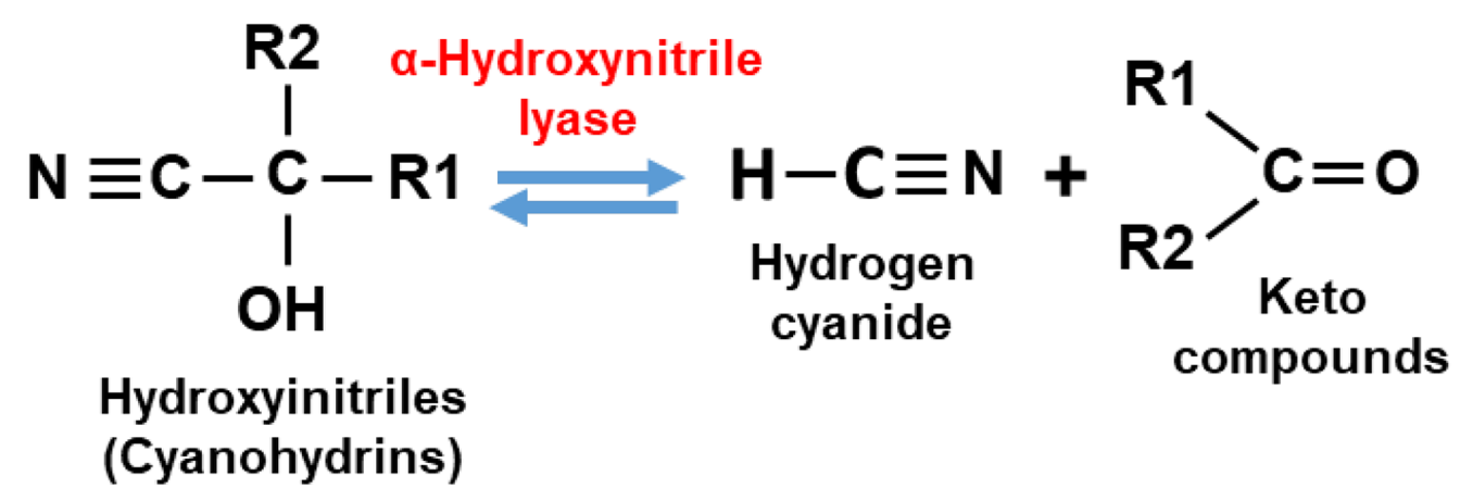
Fig. 6 Reaction of a cyanohydrin into HCN and a keto compound catalyzed by a-hydroxynitrile lyase (Boter & Diaz, 2023).
Cyanohydrin breakdown induces other byproducts such as benzaldehyde and benzoyl cyanide which are toxic compounds. Finally, benzoyl cyanide is involved with the production of hydrogen peroxide which is also highly cytotoxic (Boter & Diaz, 2023). Therefore, cyanogenic glucosides can be considered very potent and efficient due to the cascade of chemical reactions and the many toxic metabolites that are generated from that molecule.
In the Eucalyptus genus, main cyanogenic glucosides are prunasin, sambunigrin and amygdalin, displayed in Figure 7. Prunasin and sambunigrin both showcase one sugar while amygdalin has a disaccharide. Prunasin is by far the most common in the genus (Gleadow et al., 2008). Prunasin and sambunigrin are diastereoisomers while amygdalin is the diglucoside of sambunigrin. Table 1 presents a study that depicts cyanogenesis and the ratio between sambunigrin and prunasin (S:P).

Fig. 7 Cyanogenic glucosides present in Eucalyptus; 1) prunasin; 2) sambunigrin; 3) amygdalin (Gleadow et al., 2008).
Table 1 Ratio of sambunigrin to prunasin (S:P) by spectra from GLC (gas-liquid chromatography) and NMR (nuclear magnetic resonance spectroscopy) by species (Gleadow et al., 2008).
| Species | Major cyanogen | S:P GLC | S:P NMR |
|---|---|---|---|
| E.burdettiana | prunasin (1) | 0:100 | |
| E.caleyi | prunasin (1) | 8:92 | 9:91 |
| E.cylindriflora | prunasin (1) | 2:98 | 2:98 |
| E.cylindriflora | prunasin (1) | 16:84 | 15:85 |
| E.leptophleba | prunasin (1) | 0:100 | |
| E.leucoxylon | prunasin (1) | 0:100 | 22:78 |
| E.leucoxylonb | prunasin (1) | 0:100 | 0:100 |
| E.leucoxylon | prunasin (1) | 0:100 | 0:100 |
| E.megacornuta | prunasin (1) | 0:100 | 0:100 |
| E.megacornuta | prunasin (1)/sambunigrin (2) | 47:53 | 46:54 |
| E.orgadophila | prunasin (1) | 0:100 | 0:100 |
| E.orgadophila | prunasin (1) | 19:81 | |
| E.orgadophila | prunasin (1) | 24:76 | 7:93 |
| E.ovata | prunasin (1) | 0:100 | |
| E.patellaris | prunasin (1) | 0:100 | |
| E.steedmanii | prunasin (1) | 0:100 | |
| E.yarraensis | prunasin (1) | 0:100 | 0:100 |
Diversity and Ecological Perspective
Cyanogenesis in eucalyptus helps individuals grow and reproduce more effectively as their leaves, flower and fruit are considerably less targeted by herbivore or pest. Indeed, koalas that have the choice of feeding on cyanogenic and non-cyanogenic eucalyptus species will only feed on non-cyanogenic species (Pahl, 1987). Furthermore, scientists have established that cyanogenic glucosides concentration inversely correlates with leaf damage and herbivore feeding (Gleadow & Woodrow, 2002).
Cyanogenesis tends to be more strongly expressed in Eucalyptus occupying vulnerable position or fragile conditions. In fact, younger individuals of cyanogenic species such as E. cladocalyx are known to contain a higher cyanogenic glucoside concentration (Hansen et al., 2018). As younger individuals are often richer in nutrients and have less fiber, they are targeted by herbivore. Hence, producing higher levels of cyanogenic compounds during early growth provides a selective advantage and increases the likelihood of reaching reproductive maturity.
Water stress is another factor influencing the expression of cyanogenesis. Eucalyptus cladocalyx exhibits stronger cyanogenic traits when exposed to water scarcity (Woodrow et al., 2002). Such a drought-induced response reflects an adaptive mechanism to avoid predation during period of stress. Overall, Eucalyptus uses its cyanogenic polymorphism to adjust and optimize survival chances under varying environmental stress.
Cyanogenesis is not widespread in the Eucalyptus genus with only 23 cyanogenic species out of 420 described species. This only accounts for 4% of the total number of species. Although Eucalyptus subgenera Symphyomyrtus was previously thought to be the only subgenus with cyanogenic property, recent studies demonstrate two cyanogenic individuals in subgenera Eucalyptus and Corymiba respectively (Gleadow et al., 2008). This significant detail hints for convergent evolution where one mechanism is so effective that different species independently develops it to increase its fitness.
Design, Engineering and Possibility
Cyanogenesis in Eucalyptus demonstrates a nature’s design solution of controlled toxicity. By ingeniously storing cyanogenic glucoside in a separate compartment than the enzyme activating the biochemical cascade, eucalyptus ensures its own cellular safety. This principle of compartmentalized activation could guide engineered systems requiring precise control of dangerous reactions. Moreover, cyanogenic eucalyptus activates its chemical defense rapidly and effectively when need arises.
The ability of one compound generating a chain of reactions with multiple varying toxic products finds itself to be extremely relevant in this context. A set of multiple toxins can repel herbivores that developed resistance for a particular compound. Thus, it broadens the range of species it can deter using a single reactant. Such a characteristic can be significant in application of pesticides and herbivore dissuasion.
Eucalyptus proposes an example of adaptive economy by ontogenetically providing sapling higher concentrations of cyanogenic agents and producing more cyanogenic glucoside during water scarcity. That way the process of enzyme production and metabolism around cyanogenesis are only operated when the tree has a need for protection. This property brilliantly maximizes survival rates while saving heavy metabolic costs when not needed.
Overall, this cyanogenic property, among many, must be considered as a Eucalyptus’ natural design chemical solution. Eucalyptus drastically improves its survival and fitness by developing such a chemical defense allowing some species of the genus to thrive in herbivore’s territory. Moreover, beyond its ecological role, cyanogenesis represents a remarkably efficient and useful natural design – a controlled liberation mechanism that balances toxicity, economy and adaptability, offering insight into biological and biomimetic innovations. Biomimetic applications derived from Eucalyptus cyanogenesis such as biodegradable pesticide, controlled release system or safe toxic chemical storage further highlight its relevance as a natural design solution. In the intricate mechanism of Eucalyptus’ cyanogenesis lies a reminder that nature’s most efficient design often emerges from the balance between danger and control.
Chemistry of the Lignin Biopolymer
While cellulose is primarily recited as the building block of plant cell walls and extracellular matrices, the structure of plant cells, including of the Eucalyptus, can be thought of as being comprised of three primary biopolymers: cellulose, hemicellulose, and lignin (Wang et al., 2008). Cellulose is made of β-glucose monomers and is characterized by long, mostly unbranching organic chains contributing to its rigidity (Rencoret et al., 2011). Hemicellulose is made of xylose, galactose, mannose, rhamnose, and arabinose and is characterized by shorter, branched chains contributing to its flexibility and linking capabilities. Lignin is a reinforcement for the lignocellulosic matrix and provides rigidity, water-impermeability, and microbial resistance (Wen et al., 2013). It is a phenolic biomacromolecule derived from three main hydroxycinnamyl alcohols or monolignols: p-coumaryl alcohol, coniferyl alcohol, and sinapyl alcohol (Wen et al., 2013). Through free radical generation, these alcoholic monomers assemble into racemic phenylpropane macromolecules p-hydroxyphenyl (H), guaiacyl (G), and syringyl (S) via free radical coupling, as seen in Figure 8.

Fig. 8 Lignin phenylpropanoid units. The three primary monolignols are characterized by a benzene ring with a unique number of methoxyl groups. The monolignols assemble into phenylpropane units with related chemical characteristics (Wen et al., 2016).
S has two methoxyl groups, G has one methoxyl group, and H has no methoxyl groups (Wang et al., 2017). The phenylpropane units are primarily linked through aryl ether (β-O-4) bonds where the β-carbon of one monomer is connected via an ether oxygen to the fourth carbon of another monomer’s aromatic ring. Zooming out, the lignin and carbohydrate lignocellulosic matrix is formed by different chemical linkages like phenyl glycosides, benzyl ethers, and γ-esters. The hydroxycinnamyl alcohols impart rigidity to cell walls to strengthen xylem and phloem tissue fiber cells and impart water-impermeability through their hydrophobic properties (Hawkins & Boudet, 2003). In Eucalypti, this contributes to improved water retention and hydraulic efficiency.
Lignin’s Contribution to Biomass Recalcitrance
The same characteristics that make lignin effective as one of a Eucalyptus’ biopolymers serve as obstacles in herbivore feeding and in anthropogenic pulping and paper-making industries. The lignin and cellulose interactions are varied and complex, meaning that the lignocellulosic matrix contributes hugely to biomass recalcitrance (Li et al., 2016). Biologically, recalcitrance is the plant’s manner of growing upwards and resisting degradation of its cell wall polysaccharides (Ferraz et al., 2014). Lignocellulosic biomass is recalcitrant to enzymatic digestions: lignin reduces cellulose hydrolysis by forming a physical barrier that impedes enzyme access to cellulose, and it preventatively binds to cellulolytic enzymes. Specifically, lignin irreversibly causes adsorption of cellulase enzymes found in herbivore digestive systems, as seen in Figure 9.

Fig. 9 Mechanism of lignin reduction of cellulose hydrolysis. Lignin can form a physical barrier to prevent cellulase-cellulose complex or bind and deactivate the enzyme, preventing cellulose depolymerization (Yao, et al., 2022).
Consequently, plant tissues high in lignin concentration, like Eucalyptus leaves, are resilient to herbivores. Industrially, recalcitrance is a plant’s resistance to release their sugars for fermentation and is a major barrier to efficient, economical production of biofuels (Gilna et al., 2017). In this way, lignin’s chemical properties allow it to serve as a physical protection against mechanical degradation (whether by humans or possums, for example) and is a form of structural determination and resilience.
Lignin as a Defense Mechanism
Unlike animals, plants don’t have an adaptive immune system, so, upon internal breaches, they rely on each cell to respond to and defend against microbial and pest attacks through recognition, signaling, and defensive product production (Naidoo et al., 2014). Eucalypti must have defenses against pests like soft, brown, and white rot fungi, myrtle rust Puccinia psidii, and insect pest Leptocybe invasa in order to survive (Naidoo et al., 2014). The first defense against pests are the physical barriers of Eucalypti outer tissues: bark, pectin, and the lignin in plant cell walls. When the first defense is overcome, cell responsive mechanisms are initiated. The mechanisms include rapid production of ROS at the pathogen invasion site (oxidative burst), calcium ion signaling pathways to stimulate transcription of pathogenesis or apoptosis genes, phytohormone cascades, and development of papillae “scabs” and barrier zones in wounded or distal cell regions (Naidoo et al., 2014). When a pathogen inflicts physical harm to Eucalypti cells, cell walls near the wound site are reinforced by defense lignin which has been differentiated from developmental lignin because of different biosynthesis transduction pathways, gene regulation, and enzymatic activities (Hawkins & Boudet, 2003). In their 2003 study, Hawkins & Boudet histochemically analyzed the lignin reinforced barrier zones of Eucalyptus gunnii plants after wounds were inflicted. Regions of the plants were decapitated with sterile razor blades, and the wounded tissue was excised on alternating days post-injury. The tissue was then analyzed: UV light was used to identify phenolic material due to its autofluorescence, phloroglucinol-HCl was used to reveal lignin, and the Maule reaction was used to identify relative S lignin profiles via a reaction with KMnO4, HCl, and NaOH with the two methoxyl groups of S lignin. The results were distinct, as seen in Figure 10 (Hawkins & Boudet, 2003).

Fig. 10 Histochemical analysis of E. gunnii wounded tissue as viewed transversely (a-d) and longitudinally (e-f) for control (a) and wounded samples (b-f). (a) Positive red response to Maule reaction in mature secondary xylem (s) and phloem (p) indicating S lignin; negative brown response in primary xylem (px) and developing secondary xylem (sx). (b) Xylem cell blockages (b), cortex (c) and vascular cambium cell walls (v) fluoresce under UV light indicating phenolic material presence. (c) Cortex cell walls (c) appear red indicating lignin material via phloroglucinol-HCl reaction. (d) (c) and vascular cambium cell walls (v) appear brown indicating negative response to Maule reaction and low S lignin presence. (e) Cortex cell walls (c) appear red indicating lignin presence. (f) Pith cell walls (p) fluoresce under UV light indicating phenolic material presence (Hawkins & Boudet, 2003).
Within five to seven days, a well-sealed barrier formed around the wound sight. The cell walls in newly formed vessels reacted positively with the Maule reagent in the Maule reaction, indicating that S lignin was produced in immature cells as opposed to its usual production in mature cells. However, E. gunnii developmental (mature) tissue has a higher S/G lignin ratio, meaning that defense lignin produced in mature cells during wound response is abnormally poor in S units compared to developmental lignin. This indicates that the E. gunnii defensive production of lignin spurred an abnormal enrichment of S lignin in immature cells and an abnormal depletion of S lignin in mature cells. Additionally, enzymatic analyses of the regions showed that coniferyl alcohol dehydrogenase (CAD) and sinapyl alcohol dehydrogenase (SAD) activity increases as a response to wounding which is different than typically observed hydroxycinnamyl alcohol dehydrogenase activity. This indicates a difference in the chemical composition of defense lignin compared to developmental lignin. E. gunnii was triggered to initiate a different lignin production pathway, meaning that lignin’s chemical composition is specific to its purpose in Eucalyptus cells and is a tool that can be used for static structural support or active responses to attacks.
Metal Accumulation in Eucalyptus Plants through Phytoremediation
Phytoremediation is the process by which plants remove pollutants from the environment. It solves the urgent need for alternative, cheap, and efficient methods to clean up heavily contaminated industrial areas (Peuke, 2005). Specifically, phytoremediation uses wild or genetically modified plants (GMPs) to extract a wide range of heavy metals from the soil and accumulate them in the biomass of plants.
Usually, once the plants have accumulated the unwanted metals from the soil, then harvests occur to remove the plants themselves with the pollutants. These harvested plants are used efficiently in alternative processes such as burning of energy production, or simply for disposal. The advantages of phytoremediation are cheap maintenance costs, no additional carbon dioxide released if it is a carbon-dioxide neutral technology, and the possibility of potential profits by using the final biomass for heat or energy production. The disadvantage of phytoremediation is its slow process, which requires often decades to accumulate enough metal (Peuke, 2005).
Chelating compounds, with cysteine-rich polypeptides, exploit their property of binding heavy metals to the thiol-groups of proteins. This binding allows for detoxification since leaving the heavy metals on their own can have toxic effects on the surrounding environment. Glutathione (GSH) synthesizes phytochelatins, and its derivatives in the presence of heavy metals ions through the process of phytocehlatine synthase. Phytochelatins are a good indicator of heavy-metal accumulation in plants, as they were found in yeast, algae, and lower and higher plants after exposure to cadmium or copper (Cobbett, 2000). As seen in Figure 11, once the phytochelatins are synthesized, they form ligand complexes with these metals, which are then brought to the vacuole for detoxification.

Fig. 11 Plant cell mechanisms to detoxify heavy metals by glutathione (Peuke, 2005).
Certain plants have this rare ability to tolerate large concentrations of heavy metals. It is common in particular groups of plants with hyperaccumulating or metal-tolerant capabilities such as Silene vulgaris, Thlaspi caerulescens and Alyssum lesbiacum, whose common names are bladder campion, alpine pennycress, and sweet alyssum, respectively. Their metal accumulation includes essential nutrients such as copper, iron, zinc, and selenium, as well as non-essential metals such as cadmium, mercury, lead, and aluminum. Oftentimes, metal-accumulating plants have 100 to 1000 times higher metal concentration in their shoots, which is a great mechanism to use to detoxify heavy-metal concentrated soil areas (Peuke, 2005).
Factors of Heavy Metal Accumulation Abilities
The accumulation of metals in certain plants is a time-dependent process, which often takes several years to occur (Peuke, 2005). Certain plants are faster in accumulating and detoxifying metals from the soil than other plants, and the time it takes usually depends on two factors. The first factor would be the total amount of biomass that these plants produce. The second factor would be the plants’ specific metal bioconcentration factor, which is the ratio of metal concentration in the shoot tissue to the soil. Hyperaccumulators aside, most plants have this ratio equal to values less than 1, meaning it would take longer than a human lifespan to reduce the contamination in the soil by 50%. Thus, to accelerate the accumulation of metals in the plant from the soil, there must be either the use of plants with large biomass production or with a metal bioconcentration factor much greater than 1, or a combination of both species (Peuke, 2005).
Phytoextraction Capabilities of Different Eucalyptus Species
The Eucalyptus trees were studied to determine the levels of metal accumulation throughout their different species. They were chosen since they are known to be a fast-growing, short-rotation species. The four species studied were the Eucalyptus tereticornis, Eucalyptus camaldulensis (ECam), Eucalyptus globulus and Eucalyptus citriodora (ECit), and the two toxic metals used were cadmium and lead. It is observed that Eucalyptus is a possible solution to minimize heavy metal contamination in polluted soil and water sources. The main results from the study indicated that as the concentration of heavy metals increased in the soil, so did their accumulation in all of the eucalyptus shoots. Furthermore, lead was favored compared to cadmium as the metal with higher concentrations found in the root for all 4 species. ECit had the highest accumulation in its root, while ECam had the lowest accumulation. As seen in Table 2, ECit had the highest accumulation in its root at 31.68± 0.191 mg kg−1, while ECam had the lowest accumulation at 13.49± 0.023 mg kg−1 (Patil, et al., 2023).
Table 2 Phytoextraction concentration (ppm) in the root of 4 Eucalyptus species (Patil et al., 2023)
| Phytoextraction root (mg kg-1) | |||||
|---|---|---|---|---|---|
| Metal | Treatments | E1 | E2 | E3 | E4 |
| Cadmium | T1 | 6.64 ± 0.012 | 4.37 ± 0.067 | 6.35 ± 0.090 | 10.39 ± 0.073 |
| (M1) | T2 | 8.59 ± 0.045 | 5.33 ± 0.009 | 7.7 ± 0.084 | 12.62 ± 0.129 |
| T3 | 10.56 ± 0.006 | 6.71 ± 0.113 | 8.72 ± 0.064 | 17.09 ± 0.134 | |
| Lead | T1 | 17.19 ± 0.262 | 9.22 ± 0.006 | 14.33 ± 0.13 | 19.59 ± 0.038 |
| (M2) | T2 | 18.5 ± 0.023 | 10.31 ± 0.058 | 16.21 ± 0.33 | 21.88 ± 0.216 |
| T3 | 26.33 ± 0.215 | 13.49 ± 0.023 | 21.05 ± 0.003 | 31.68 ± 0.191 | |
| ANOVA test of significance at 0.05 | E=0.202** | T = 0.175** | M=0.143** | ExMxT=0.495** | |
| Note: DAS, days after sowing; E1—E. tereticornis, E2—E. camaldulensis, E3—E. globulus, E4—E. citriodora; M1T1—cadmium 25 ppm, M1T2—cadmium 50 ppm, M1T3—cadmium 100 ppm, M2T1—lead 100 ppm, M2T2—lead 125 ppm, M2T3—lead 250 ppm | |||||
In addition, since most of the plants in this study were growing at the same time, their roots which are constantly changing operate as a barrier, which will prevent metals from moving to the above-ground parts of the plant and thus reducing the level of metal accumulation upwards and increasing its concentration near the roots. Based on the data, ECit had the ability to extract metals at greater hazard metal concentration levels and because it grew faster, it was able tolerate higher levels of toxicity (Patil, 2024). Among all the species, Eucalyptus camaldulensis had the lowest accumulation rate.
Decontamination of Soil and Water through Eucalyptus Roots
The eucalyptus trees have been designed to biologically solve the problem of heavy metal contamination in soil and water sources. The roots of Eucalyptus species act as biological filters, trapping the heavy metal ions within their roots. Moreover, due to their ability to accumulate heavy metals in their roots, which play a role in mechanisms involving synthesizes of GSH and PCs, they are able to utilize these ions in chemical processes and repurpose them for bioenergy production. Over time, through multiples planting and harvesting cycles, the use of large quantities of Eucalyptus trees in high heavy-metal concentrated areas can lead to decontamination of the area, which will overall reduce the concentration of toxic metals entering the food chain.
Conclusion
Eucalyptus’ structural, defensive, and survivalist components are determined by their chemical properties. In an environment abundant with insects, fungi, and microbes targeting Eucalyptus leaves and outer tissue, terpenoid essential oils repel herbivores and insects through enzyme inhibition and antifeedant vapors. Although the toxicity of these molecules is notable, their ability to volatize into acrid vapors fulfills a multimodal defense system: the herbivores are deterred through both sensory experience and internal toxicity, a property that can be harnessed for antimicrobial materials (bandages, packaging) or pest deterrents that only activate when pests contact the crop. Similarly, Eucalypti can harness selective toxicity through activation of cyanogenic glucosides, deterring herbivores and feeding. The compartmentalized activation of harmful HCN is a key design solution: the Eucalyptus can sequester and produce a deadly compound, turning it into a weapon through specified activation. The HCN itself isn’t attacking the herbivores; it’s the process of activation that turns the HCN into Eucalyptus’ tool. Drug delivery systems can benefit from this solution through adapted treatments that are activated at essential times to overcome the human body’s defense systems, like mucosal membranes. Internally, lignin serves as a structural foundation with its rigid qualities and as a defensive shield, forming reinforced tissue around wounds to prevent further harm. The duality of mundane and responsive function is an energy-saving solution to the Eucalyptus system and can be harnessed in products that would benefit from localized repair upon damage. Building composites like reinforced concrete could utilize a capsule repair system where when cracked, a resin and catalyst would mix to reinforce the damaged region. Eucalyptus chemical processes also lead to environmental impacts, like how the volatile oils are allelopathic and the roots of the Eucalyptus can detoxify water sources from heavy metals. The reuse of toxic metals as components of future energy production highlights the creative and resilient survival properties that allow the Eucalyptus to be dominant in their ecosystems. These characteristics can be directly applied to environmentally engineered rain gardens or artificial marshes constructed in cities: column or mat structures that mimic the Eucalyptus roots can act as filters to toxic metal runoff and other pollutants. The complex and versatile methods of survival and prospering of the Eucalyptus are owed to the distinct chemical characteristics of its essential molecules.
References
References
Andualem, A.M., Aragaw, M.W., Molla, A.E. et al. (2024). Allelopathic effects of leaf extracts of Eucalyptus camaldulensis Dehnh. on morphological, physiological, and yield traits of Ethiopian wheat (Triticum durum L.) cultivars. BMC Plant Biol 24, 1138. https://doi.org/10.1186/s12870-024-05832-9
Belaid, S., Gonzalez‐Coloma, A., Andres, M. F., Elfalleh, W., Idoudi, S., Romdhane, M., & Saadaoui, E. (2025). Exploring chemical profiles, antifeedant, nematicidal, and phytotoxic potentials of seven essential oils from eucalyptus species. Chemistry & Biodiversity, 22(6). https://doi.org/10.1002/cbdv.202402960
Boter, M., & Diaz, I. (2023). Cyanogenesis, a Plant Defence Strategy against Herbivores. Int J Mol Sci, 24(8). https://doi.org/10.3390/ijms24086982
Chu, C., Mortimer, P. E., Wang, H., Wang, Y., Liu, X., & Yu, S. (2014). Allelopathic effects of eucalyptus on native and introduced tree species. Forest Ecology and Management, 323, 79–84. https://doi.org/10.1016/j.foreco.2014.03.004
Cobbett C. S. (2000). Phytochelatins and their roles in heavy metal detoxification. Plant physiology, 123(3), 825–832. https://doi.org/10.1104/pp.123.3.825
Darben, T., Cominos, B., & Lee, C. T. (1998). Topical eucalyptus oil poisoning. The Australasian journal of dermatology, 39(4), 265–267. https://doi.org/10.1111/j.1440-0960.1998.tb01488.x
Dellacassa, E., & Moyna, P. (1992). Eucalyptus leaf oils, use, chemistry, distillation and marketing, edited by D. J. Boland, J. J. Brophy and A. P. N. House, Inkata Press, Melbourne, 1991. A$65.00. ISBN 0–85066–423–3. no. of pages: 252. Flavour and Fragrance Journal, 7(2), 105–105. https://doi.org/10.1002/ffj.2730070209
del Moral, Roger & Muller, Cornelius. (1970). The Allelopathic Effects of Eucalyptus camaldulensis. The American Midland Naturalist. 83. 254-282. 10.2307/2424020. https://doi.org/10.2307/2424020
Dumanović, J., Nepovimova, E., Natić, M., Kuča, K., & Jaćević, V. (2021). The significance of reactive oxygen species and antioxidant defense system in plants: A concise overview. Frontiers in Plant Science, 11. https://doi.org/10.3389/fpls.2020.552969
Elangovan, S., & Mudgil, P. (2023). Antibacterial Properties of Eucalyptus globulus Essential Oil against MRSA: A Systematic Review. Antibiotics (Basel, Switzerland), 12(3), 474. https://doi.org/10.3390/antibiotics12030474
Ferraz, A., Costa, T.H.F., Siqueira, G., Milagres, A.M.F. (2014). Mapping of Cell Wall Components in Lignified Biomass as a Tool to Understand Recalcitrance. In: da Silva, S., Chandel, A. (eds) Biofuels in Brazil. Springer, Cham. https://doi.org/10.1007/978-3-319-05020-1_9
Gilna, P., Lynd, L. R., Mohnen, D., Davis, M. F., & Davison, B. H. (2017). Progress in understanding and overcoming biomass recalcitrance: a BioEnergy Science Center (BESC) perspective. Biotechnology for biofuels, 10, 285. https://doi.org/10.1186/s13068-017-0971-1
Gleadow, R. M., Haburjak, J., Dunn, J. E., Conn, M. E., & Conn, E. E. (2008). Frequency and distribution of cyanogenic glycosides in Eucalyptus L'Herit. Phytochemistry, 69(9), 1870-1874. https://doi.org/10.1016/j.phytochem.2008.03.018
Gleadow, R. M., & Woodrow, I. E. (2002). Defense chemistry of cyanogenic Eucalyptus cladocalyx seedlings is affected by water supply. Tree Physiol, 22(13), 939-945. https://doi.org/10.1093/treephys/22.13.939
Gullón, P., Gullón, B., Astray, G., Munekata, P. E. S., Pateiro, M., & Lorenzo, J. M. (2020). Value-Added Compound Recovery from Invasive Forest for Biofunctional Applications: Eucalyptus Species as a Case Study. Molecules (Basel, Switzerland), 25(18), 4227. https://doi.org/10.3390/molecules25184227
Hansen, C. C., Sorensen, M., Veiga, T. A. M., Zibrandtsen, J. F. S., Heskes, A. M., Olsen, C. E., Boughton, B. A., Moller, B. L., & Neilson, E. H. J. (2018). Reconfigured Cyanogenic Glucoside Biosynthesis in Eucalyptus cladocalyx Involves a Cytochrome P450 CYP706C55. Plant Physiol, 178(3), 1081-1095. https://doi.org/10.1104/pp.18.00998
Hawkins, S., & Boudet, A. (2003). ‘defence lignin’ and hydroxycinnamyl alcohol dehydrogenase activities in wounded eucalyptus gunnii. Forest Pathology, 33(5), 339–352. https://doi.org/10.1046/j.1439-0329.2003.00341.x
Li, H.-Y., Wang, C.-Z., Chen, X., Cao, X.-F., Sun, S.-N., & Sun, R.-C. (2016). Structural elucidation of eucalyptus lignin and its dynamic changes in the cell walls during an integrated process of ionic liquids and successive alkali treatments. Bioresource Technology, 222, 175–181. https://doi.org/10.1016/j.biortech.2016.09.111
Li, Zhao-Hui & Wang, Qiang & Ruan, Xiao & Cunde, Pan & Jiang, De-An. (2010). Phenolics and Plant Allelopathy. Molecules. 15. 8933-8952. 10.3390/molecules15128933. https://doi.org/10.3390/molecules15128933
Kim, K. Y., Seo, H. J., Min, S. S., Park, M., & Seol, G. H. (2014a). The effect of 1,8‐Cineole inhalation on preoperative anxiety: A randomized clinical trial. Evidence-Based Complementary and Alternative Medicine, 2014(1). https://doi.org/10.1155/2014/820126
McKemy DD. TRPM8: The Cold and Menthol Receptor. In: Liedtke WB, Heller S, editors. TRP Ion Channel Function in Sensory Transduction and Cellular Signaling Cascades. Boca Raton (FL): CRC Press/Taylor & Francis; 2007. Chapter 13. Available from: https://www.ncbi.nlm.nih.gov/books/NBK5238/
Mineur, Y. S., & Picciotto, M. R. (2021). The role of acetylcholine in negative encoding bias: Too much of a good thing?. The European journal of neuroscience, 53(1), 114–125. https://doi.org/10.1111/ejn.14641
Morris, P. F., & Pescott, E. E. (1944). Poisoning by eucalypts. Via. Nat, 60(31), Article 19440701171. https://trove.nla.gov.au/newspaper/article/84053858
Müller, F., & Silber, S. (2012). Polysiloxanes. Polymer Science: A Comprehensive Reference, 443–451. https://doi.org/10.1016/b978-0-444-53349-4.00276-4
Naidoo, S., Kulheim, C., Zwart, L., Mangwanda, R., Oates, C. N., Visser, E. A., Wilken, F. E., Mamni, T. B., & Myburg, A. A. (2014). Uncovering the defence responses of eucalyptus to pests and pathogens in the genomics age. Tree Physiology, 34(9), 931–943. https://doi.org/10.1093/treephys/tpu075
National Center for Biotechnology Information (2025). PubChem Compound Summary for CID 2758, 1,8-Cineole. Retrieved October 16, 2025 from https://pubchem.ncbi.nlm.nih.gov/compound/1_8-Cineole.
Naudé, Y., Makuwa, R., & Maharaj, V. (2016). Investigating volatile compounds in the vapour phase of (1) a hot water infusion of rhizomes, and of (2) rhizomes of Siphonochilus aethiopicus using head space solid phase microextraction and gas chromatography with time of flight mass spectrometry. South African Journal of Botany, 106, 144-148. https://doi.org/10.1016/j.sajb.2016.07.006
Pahl, L. I. (1987). Feeding-Behavior and Diet of the Common Ringtail Possum, Pseudocheirus-Peregrinus, in Eucalyptus Woodlands and Leptospermum Thickets in Southern Victoria. Australian Journal of Zoology, 35(5). https://doi.org/10.1071/zo9870487
Patil G., Divya M.P., Shah, P., & Maitry, A. (2023). Metal accumulation ability of different Eucalyptus species at the early stage. Forestist, 74(1), 26-34.
https://forestist.org/Content/files/sayilar/260/FRSTST_20220078_nlm_new_indd_.pdf
Peuke, A. D., & Rennenberg, H. (2005). Phytoremediation. EMBO reports, 6(6), 497–501. https://doi.org/10.1038/sj.embor.7400445
Rants'o, T. A., Koekemoer, L. L., Panayides, J. L., & van Zyl, R. L. (2022). Potential of Essential Oil-Based Anticholinesterase Insecticides against Anopheles Vectors: A Review. Molecules (Basel, Switzerland), 27(20), 7026. https://doi.org/10.3390/molecules27207026
Rencoret, J. (2000). Structural characterization of milled wood lignins from different eucalypt species. Holzforschung. https://doi.org/10.1515/HF.2008.096
Rencoret, J., Gutiérrez, A., Nieto, L., Jiménez-Barbero, J., Faulds, C. B., Kim, H., Ralph, J., Martínez, A. T., & Del Río, J. C. (2011). Lignin composition and structure in young versus adult Eucalyptus globulus plants. Plant physiology, 155(2), 667–682. https://doi.org/10.1104/pp.110.167254
Sasikumar, K. & Vijayalakshmi, C. & Parthiban, K.T.. (2002). Allelopathic effects of Eucalyptus on blackgram (Phaseolus mungo L.). Allelopathy Journal. 9. 205-214. https://www.researchgate.net/journal/Allelopathy-Journal-0973-5046?_tp=eyJjb250ZXh0Ijp7ImZpcnN0UGFnZSI6Il9kaXJlY3QiLCJwYWdlIjoicHVibGljYXRpb24ifX0
Wang, H.-M., Wang, B., Wen, J.-L., Yuan, T.-Q., & Sun, R.-C. (2017). Structural characteristics of lignin macromolecules from different eucalyptus species. ACS Sustainable Chemistry & Engineering, 5(12), 11618–11627. https://doi.org/10.1021/acssuschemeng.7b02970
Wen, J.-L., Sun, S.-L., Xue, B.-L., & Sun, R.-C. (2013). Recent Advances in Characterization of Lignin Polymer by Solution-State Nuclear Magnetic Resonance (NMR) Methodology. Materials, 6(1), 359-391. https://doi.org/10.3390/ma6010359
Woodrow, I. E., Slocum, D. J., & Gleadow, R. M. (2002). Influence of water stress on cyanogenic capacity in Eucalyptus cladocalyx. Funct Plant Biol, 29(1), 103-110. https://doi.org/10.1071/PP01116
Yao, L., Yang, H., Meng, Y., & Ragauskas, A. (2022). Toward a Fundamental Understanding of the Role of Lignin in the Biorefinery Process. Frontiers in Energy Research, 9. https://doi.org/10.3389/fenrg.2021.804086
Yulvianti, M., & Zidorn, C. (2021). Chemical Diversity of Plant Cyanogenic Glycosides: An Overview of Reported Natural Products. Molecules, 26(3), 719 https://doi.org/10.3390/molecules26030719