
Table of Contents
Keywords: photosynthesis, starch, nutritional composition, tannins, astringency, antimicrobial activity, chestnut blight
Abstract
Chestnut trees (Castanea) exhibit a complex set of chemical processes, allowing them to interact with their environment and sustain their productivity. This paper highlights the primary biochemical properties of chestnut trees by examining various metabolic activities and defense mechanisms employed by different species in response to their environmental interactions. Light availability is a critical factor influencing the photosynthesis rate of the chestnut tree and underlying several biochemical adaptations. Furthermore, chestnuts, with their high concentration of starch, represent a key element in the tree’s strategies for seed dispersal and germination. Moreover, tannins, a phenolic compound found in high abundance in this tree, play an important role in the tree’s constitutive defense mechanism. The analysis of the chemical interaction between the tree and the causal agent of the chestnut blight, Cryphonectria parasitica, allows a better understanding of blight resistance and enables the exploration of possible pathways toward the recovery of the American chestnut.
Introduction
The chestnut tree, belonging to the Castanea genus, is a famous tree known for its familiar nut (Fig. 1), which has played a vital role in cultures across the world for the past few centuries. It has many different species in the Northern Hemisphere, with the most prominent being the American chestnut, sweet chestnut, Chinese chestnut, and Japanese chestnut. The chestnut owes its longevity as a cuisine staple to the chemical mechanisms that have evolved to protect it. Gaining a deeper understanding of these remarkable chemicals and their role in the chestnut tree’s survival can help inspire industries and designs in engineering, such as leather tanning and formaldehyde removal (Sundaram et al., 2020).

Fig. 1. An American chestnut broken open to reveal the edible nut on the inside. The surrounding husk has many useful biochemical properties that are utilized in other industries, such as leather production (Stewart, 2018).
The genus has faced many challenges, but the most drastic effects have been on the American chestnut due to the blight in the 1800s. Caused by a fungal infection, this disease almost destroyed the country’s entire chestnut population. However, the Chinese chestnut’s unique chemistry provided a resilient front to prevent it from experiencing the same drastic effects. By comparing the differences between the two species and combining them with researched gene-editing and hypovirulent techniques, the American chestnut population has begun its restoration process (Rigling & Prospero, 2018).
Photosynthesis
Chestnuts rely on photosynthesis to obtain the necessary energy to power their metabolic processes. Photosynthesis can be divided into two stages: a light-dependent stage (Fig. 2), where the chlorophyll present on their leaves captures photons, and their energy is used to generate ATP and NADPH, and a second, non-light-dependent stage known as the Calvin cycle or dark reactions (Fig. 3), where the highly energetic products from the first step are consumed to synthesize glucose from atmospheric carbon dioxide.

Fig. 2. Diagram of the photosynthetic membrane. A) Photosystem II absorbs light, creating high-energy electrons. B) Water splits to replace the electrons, releasing oxygen (O₂). C) These electrons move down an electron transport chain, which pumps hydrogen ions (H+) into the thylakoid lumen. D) Photosystem I absorbs light, providing electrons to produce NADPH. E) The H+ ion gradient drives ATP synthesis. F) ATP and NADPH are then used to power the Calvin cycle (Adapted from Britannica, 2025).
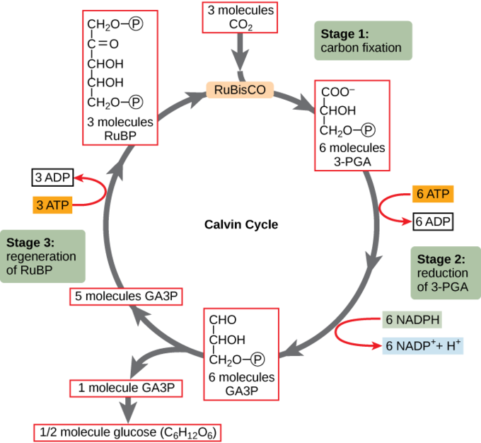
Fig. 3. Diagram of the Calvin cycle. During the first stage, CO₂ is attached to RuBP by the enzyme RuBisCO, forming 3-PGA. In the second stage, 3-PGA is converted into a three-carbon sugar (GA3P) using energy from ATP and electrons from NADPH. In the third stage, the remaining molecules are used to regenerate RuBP so the cycle can repeat. It needs to run three times to make one GA3P molecule and six times to form a glucose molecule (Carter, 2020).
The Competition for Light
Light is what powers photosynthesis and is thankfully made abundantly available by the Sun. As the first plants left aquatic environments and started to spread through land, evolve, and grow bigger, their mission became harder, given how a larger plant could get most of the light and cast a shade on smaller plants, such as young chestnut shoots.
Chestnuts are considered shade-tolerant species and have a trick up their sleeve to grow when light is scarce. They can maximize photosynthesis under low light conditions with the regulation of specific gene expression. A study conducted by Yang’s team on the Chinese chestnut, which usually grows on shaded mountain slopes, highlighted that under low-light stress the expression of the CmLhcb2.1 gene, located in the chloroplast, is upregulated. This gene is associated with an increased synthesis of chlorophyll b and of light-harvesting complex proteins (Yang et al., 2025). Since chlorophyll a content is generally higher in mature chestnut leaves, increasing the concentration level of chlorophyll b under low-light stress represents a biochemical solution that extends the range of light wavelengths absorbed by Photosystem II (PSII), thereby increasing photosynthesis efficiency (Turfan et al., 2020). This adaptation is enhanced by morphological and physiological changes, such as adjusting their specific leaf area (SLA) or leaf mass per area (LMA). In low-light or shade environments, these plants produce thinner leaves with higher SLA and lower LMA to maximize light capture per unit biomass. Meanwhile, in well-lit environments, they display thicker, denser leaves with more photosynthetic machinery to utilize the abundant energy (Proietti et al., 2000; Wang et al., 2006). American chestnut seedlings grown in shade have a significantly higher SLA than those in full sun, which allows the seedlings to support more leaf area with the same biomass (Wang et al., 2006). Sweet chestnut leaves are 26% thinner than sunlit leaves, with reduced thickness in both epidermal and mesophyll tissues (Proietti et al., 2000).
Nutritional Value
Chemical Composition of Chestnuts and Environmental Influences
For the chestnut tree to prosper in so many different environments across the Northern Hemisphere, it had to evolve a flexible nutrient absorption system. The chemical characteristics of chestnut vary depending on environmental conditions, such as altitude, latitude, and climate. Soil characteristics and precipitation rate strongly influence the quantity of nutrients available (Martínez et al., 2022; Yang et al., 2015). For instance, the chestnut mineral content depends directly on the nature of the soil, meaning that if there is a higher content of calcium in the soil, the nut will also have a great amount of calcium. However, Yang’s research team compared the minerals detected in chestnuts grown in different regions with very similar iron levels in the soil and found a significant difference in iron concentration within the chestnut. This is because trace metals influence mineral absorption differently than other nutrients, such as calcium. For example, high aluminum absorption inhibits the absorption of iron. Additionally, the form of iron (free or bound) influences how efficiently it is absorbed (Yang et al., 2015).
Starch Composition and Role in Germination
Parent chestnut trees supply their offspring with all the essential nutrients and energy that they need to grow, neatly packed within the seed. During germination, the seed goes through a series of metabolic reactions to grow, mainly by breaking down stored nutrients into usable energy. Chestnut is mostly composed of starch, with an estimated 43g of starch per 100g dry seed matter. When the seed attains the ideal temperature and moisture conditions, germination begins and causes starch hydrolysis. This means that starch is broken down by amylase into dextrin and maltose, which are then broken down into glucose by maltase (Santos et al., 2022).
Glucose is used as energy for plant growth, since carbon-carbon (C-C) and carbon-hydrogen (C-H) bonds store a high amount of potential energy due to equally shared electrons (Sharpe, 2011). The parent tree initially converted its glucose molecules into starch to store energy to give to its offspring. Additionally, starch is used by the parent tree as a backup energy source when light is not available (Pfister & Zeeman, 2016). While starch acts as an energy reserve for the tree, insufficient starch production leads to a slower growth rate. Starch is primarily formed of two types of glucose polymers: amylose and amylopectin (Fig. 4). Together, they form starch granules in which amylopectin forms the structure and amylose fills the gaps of the semi-crystalline structure built by amylopectin (Fig. 5) (Pascoli Cereda, 2023). Furthermore, chestnut not only polymerizes because it allows it to store energy in compacted form, but also because starch is insoluble in water. This means that it protects the tree from osmotic damage. Osmotic pressure is a force resulting from the intake of water into the cell to decrease the high concentration of dissolved solutes inside the cell. If glucose is not converted into starch, the cell has a high concentration of glucose dissolved, which causes a high amount of water molecules to penetrate the cell to balance out the concentrations. This causes stress that can impact growth. Therefore, by converting glucose into an insoluble form, it eliminated the issue of osmotic damage (Koroleva et al., 2002).

Fig. 4. Comparison of amylose and amylopectin structure. Amyloses consist of linear chains of glucose molecules, while amylopectin has a branched structure (Wizedemy Inc., 2025, https://www.citationmachine.net/bibliographies/3b0a506d-36ef-4a2a-9daf-76cf6585b73b)

Fig. 5. Organization of starch granules with amylopectin and amylose. This shows how amylose is embedded in branched amylopectin (Perin & Murano, 2017).
Tannins
Defense Mechanism
The chestnut tree readily attracts small mammals and insects because of its nutritious chestnuts, which helps the tree distribute its seeds through a mutualistic relationship. However, without a proper defense system, the rest of the tree (i.e., the leaves, roots, and trunk) will also be susceptible to consumption by these herbivores. Luckily, the chestnut tree is abundant in phenolic compounds called tannins, which are produced in almost every part of the tree because of their fundamental role in protecting the tree through an evolved cross-linking mechanism (Soares et al., 2020). Phenolic compounds are a wide variety of molecules containing one or more hydroxyl groups attached to at least one aromatic ring. These compounds are the most abundant secondary metabolites in all plants, meaning they are not essential for growth but help defend against pathogens and compete with neighboring species (Su et al., 2025).
There are two types of tannins found in the chestnut tree: hydrolysable and condensed tannins. As their name suggests, hydrolysable tannins can easily undergo hydrolysis and are classified based on their products. They contain a sugar core with phenolic acids attached to it by ester bonds. The main type of hydrolysable tannins found in chestnuts is ellagitannins, which form ellagic acid, gallic acid, and glucose after reacting with water (Pizzi, 2019). The four ellagitannins that are most abundant in chestnut tannin are castalagin (53%), vescalagin (35%), castalin (3%), and vescalins (8%) (Fig. 6). Condensed tannins are polymers composed of a flavan-3-ol subunit without a sugar core (Fig. 7), making them resistant to hydrolysis. They are also present in chestnuts, but in much lower proportions, and are mostly credited for their antioxidant properties (Su et al., 2025).

Fig. 6. The four main ellagitannins (a type of hydrolysable tannin) found in chestnuts and chestnut trees. The most abundant have almost identical structures and are A) castalagin (R1 = OH, R2 = H) and vascalagin (R1 = H, R2 = OH), while the least abundant are B) castalin (R1 = OH, R2 = H) and vescalin (R1 = H, R2 = OH). The alkane chain seen in the center of molecule A and the top of molecule B is the sugar core. Phenolic acids are attached to the core through ester linkages, which are classified by their aromatic ring and hydroxyl groups. These acids get disconnected from the sugar core through hydrolysis at the ester linkages (Štumpf et al., 2023).

Fig. 7. Diagram of a condensed tannin. The flavan-3-ol subunit is seen in the brackets and is repeated to create a polymer. This polymer can be branched (e.g., the unit attached on the bottom left is a branch) or linear. Unlike the hydrolysable tannins, there is no sugar component involved (Moon et al., 2024).
Tannins are classified as a unique group of phenolic compounds that can bind to proteins. This special property gives tannins their characteristic astringent taste. Astringency means “to bind” and is classified as an unpleasant, dry, puckering sensation in the oral cavity, commonly experienced when eating an unripe banana. According to the mechanism first described by Bate-Smith in 1954, the perception of astringency can be described by the precipitation of saliva proteins as they react with tannins. In 1978, Hagerman and Butler expanded this theory to emphasize the role of proline-rich proteins as the highest interactants with tannins, therefore playing the strongest role in astringency (Soares et al., 2020). These proteins are highly abundant in mammalian saliva, making this strategic defense mechanism particularly relevant to the chestnut tree’s protection against squirrels and rodents (McArthur et al., 1995). Proline is a unique amino acid due to its ring structure (Fig. 8), which prevents the amino acid from folding into alpha helices or beta-pleated sheets. This prevents a high degree of folding in proline-rich proteins, which contain 25-42% proline within their amino acid composition, and therefore, it is easier for tannins to bind to them (Soares et al., 2020).

Fig. 8. Diagram of proline, a hydrophobic amino acid with a unique ring structure. Proline is the only amino acid that connects to the protein backbone twice and prevents secondary folding from occurring by restricting the rotation of the peptide chain (Umumararungu et al., 2024).
When tannins enter the mouth of a mammal, they begin their astringent mechanism by surrounding the protein and attaching to all the available binding sites. The proteins are not specific to a certain type of tannin, as the proline ring can form hydrophobic interactions with the gallic acids from hydrolysable tannins and flavan-3-ol units of condensed tannins. Hydrogen bonding occurs between the tannins and the protein backbone to stabilize the connection. As more tannins are added, one tannin molecule can bind to two or more proteins at once, which cross-links them together to form clusters called colloidal complexes. As these clusters get large enough, they will precipitate out of the saliva (Fig. 9). This reduces the viscosity of the saliva and adheres to the mucosa, forming an unpleasant, sticky, and dry residue in the mouth, which draws predators away from the roots, trunk, and leaves of the tree (Soares et al., 2020).

Fig. 9. The cross-linking mechanism of tannins precipitates proline-rich proteins in saliva, creating an astringent taste. A) The tannins surround the protein and bind through hydrophobic interactions with the proline ring and hydrogen bonding with the protein backbone. Since every phenolic acid (in hydrolysable tannins) or flavan-3-ol unit (in condensed tannins) can bind to a protein, one tannin molecule can bind to multiple proteins and cross-link them into a colloidal complex, which will precipitate out of the solution. B) The clusters adhere to saliva and form a sticky residue. C) Taste and mechanoreceptors perceive the uncomfortable sensation and send an electric signal to the brain to associate tannins with an unpalatable taste (Soares et al., 2020).
Tannins can use the same mechanism to exhibit antibacterial and antiviral behaviour. The mitochondria in fungi, insects, and plants are vital to the organisms because of their role in cellular respiration, in which an electron transport chain on their inner membrane completes the oxidation of glucose to form energy in the body. Tannins can cross-link these proteins to inhibit oxidative phosphorylation. As well, tannins can inhibit cellulase, pectinase, and xylanase, which are three enzymes produced by fungi and bacteria to break down cell wall components (cellulose, pectin, and xylan, respectively) into monomers. Since the organisms use this process to create energy, denaturing these proteins can lead to death. They can also inhibit peroxidase, an enzyme used to detoxify reactive oxygen species (ROS). These unstable molecules, which include the superoxide anion (O2-) and hydrogen peroxide (H2O2), are formed in the mitochondria when electrons leak prematurely from protein complexes in the electron transport chain. They react with oxygen to form unstable radicals that can damage cells by removing electrons from molecules in the organisms (e.g., lipids, proteins, DNA), triggering harmful chain reactions. Organisms with high concentrations of these toxins cannot remove them without peroxidase, demonstrating how tannins inhibit these vital proteins helps protect the tree from harmful pathogens (Scalbert, 1991).
Although tannins can increase the activity of ROS in other organisms, they can also be used as an antioxidant in the tree itself. When the tree experiences large environmental stresses, such as drought or UV exposure, the tree experiences a surge in ROS. This could lead to oxidative stress, which occurs when there is an imbalance between the production of ROS and the body’s ability to fight them off with antioxidants. Luckily, tannins can act as antioxidants by donating a hydrogen atom from their phenolic hydroxyl groups in a homolytic cleavage at the O-H bond. This adds an electron to the radical and stabilizes it, while leaving a new radical behind on the tannin. The aromatic structure of the tannin can delocalize this unpaired electron and create a stable radical to avoid further reactions. This helps the chestnut tree sustain harsh conditions and continue to grow (Amarowicz, 2007).
Leather and Cotton Production
Over the past few centuries, tannins have been used to tan leather by immersing skins in water with tannin-rich barks (Pizzi, 2019). The Mehrgarh civilization, which inhabited modern-day Pakistan from 7000 to 2600 BCE, is the first to be credited with dying animal skin using tannins from trees. The culture discovered that soaking their skins in tannins created a durable leather to protect their feet from the harsh ground and transport their food (Morgan, 2020). However, the specific use of chestnut as a tannin source was first commercialized in the middle of the 19th century, when chestnut tannin replaced oak chips to tan the leather in 28 days instead of one year, and became standard due to the tannins’ additional strengthening properties. The phenols of the tannin irreversibly bind to peptides in collagen and other proteins contained in the animal skins, which inhibits proteolytic enzymes that could otherwise break down the material. Compared to other woods, chestnut tannin creates the strongest and most chemically stable leather. It is also a desirable alternative to synthetic tannins due to its environmentally friendly nature. However, this makes it the most expensive tannin for commercial use (Ciriminna et al., 2024).
Chestnut tannins have also been used in cotton production, specifically to reduce formaldehyde (CH2O) traces from the material (Fig. 10). Formaldehyde is a colorless gas that can be identified by its strong, pungent odor. It is good at penetrating and damaging biological structures, and prolonged exposure can result in modifications known as chemical fixation. Humans are primarily exposed to the compound through common household items, such as furniture, decorations, textiles, plastics, and organic paints. It is very commonly found in cotton fabrics, as they are treated with formaldehyde for wrinkle prevention. Currently, the main methods for removing the compound include physical adsorption, membrane separation, photocatalytic oxidation, and chemical treatment. These methods are all successful but have various economic and environmental drawbacks, and chestnut tannins can be an efficient and green alternative to eliminate formaldehyde from indoor environments (Su et al., 2025).
When the primary ellagitannins in chestnut, castalagin and vascalagin, undergo hydrolysis, they form gallic and ellagic acid (Fig. 10). When bonded to a cotton fabric, these products can facilitate the adsorption of formaldehyde into the cotton.[1] To convert the cotton sample into an adsorption medium, it is first immersed in an aqueous solution of tannin extract and 1,4-butanediol diglycidyl ether (BDDE), which acts as a cross-linking agent and joins tannins together through covalent bonds. This differs from the natural cross-linking action of tannins in the chestnut tree, where the tannin cross-links proteins together through intermolecular forces. Then, the fabric is added to a new solution of poly diallyl dimethylammonium chloride, also known as PDMDAAC, whose positive charges counteract the negative charges of chestnut tannins in solution, which form from the weakly acidic hydroxyl groups. Through electrostatic interactions between the tannins and PDMDAAC, multilayer assembly occurs on the fabric surface and creates a layered structure on the cotton fabric (Su et al., 2025).

Fig. 10. The main products of hydrolysis of a castalagin or vescalagin. A water molecule reacts with each ester bond to form glucose (not pictured), a gallic acid, and two ellagic acid molecules (Su et al., 2025).
This process converts the cotton fabric into an adsorption medium. When the fabric is treated with a formaldehyde solution for wrinkle prevention, the formaldehyde will evaporate into the air, and the tannins will adsorb the molecules back into the cotton to stabilize them (Su et al., 2025).
Sensory Characteristics
Chestnuts rely on mutualistic relationships with seed-hoarders for seed dispersal. To attract these animals and ensure that most of their seeds have the chance to germinate and grow, chestnuts have adapted design solutions to make their seeds appealing enough to be cached, yet not so appealing that they are entirely consumed. The edible kernel inside the chestnut tends to be low in tannins, which is why mammals prefer them to any other part of the chestnut tree (Sundaram et al., 2020).
Chestnuts achieve this through distinct physical and chemical features that indicate to seed-hoarders whether a seed is dormant or germinating. A seed is considered dormant when it is inactive because of the absence of essential conditions required for germination, such as in the winter when water is frozen and light is scarce (Gubler et al., 2005).
Eastern Grey Squirrels have a mutualistic relationship with chestnuts. They show a preference of consuming germinating seeds earlier than dormant seeds. This is because germinating seeds perish quickly, so caching them would not be beneficial for the squirrel. Additionally, their nutritional value decreases as the seed germinates, since the taproot, the main central root, uses the nutrients to grow. However, since dormant seeds maintain their nutrients for longer periods of time, seed-hoarders tend to cache them for later. One of the features that the chestnut has adapted to allow distinguishing the state of the seed is the pericarp, the protective outer layer of the shell, which increases water-repellent polymers in the cell walls, such as suberin, and lignin, to become thicker during dormancy. As the seed germinates, this waxy pellicle slowly degrades and becomes more porous, releasing volatile fatty acid esters that squirrels can smell (Sundaram et al., 2020).
Chestnuts’ sensory characteristics can be explained by nutrient composition. The color of the outside nut, the pellicle, tends to be reddish to tan with varying glossiness (Fig. 11). Additionally, the ease to peel the chestnut is directly influenced by the tannin content present in the edible part of the fruit (Fig. 12) (Zhang et al., 2018).

Fig. 11. Chinese chestnut from nine chestnut variety trees grown under similar conditions have size and color variations. The varieties are as follows: A) Variety 3113, B) Benlizi, C) Dahongpao, D) Erqingzao, E) Heishanzhai 7, F) Mi 5, G) Mi 6, H) Yangguang, I) Yanshanhong (Zhang et al., 2018).

Fig. 12. View of an opening chestnut burr containing edible nuts, modified from Chestnut Growers, Inc. A) The burr is the green-yellow spikes surrounding the nuts. B) Three edible chestnuts. (Adapted from Chestnut Growers, Inc, 2021, https://chestnutgrowersinc.com/compare-w-horsechestnuts/ ).
Furthermore, the yellow color intensity of the nut is associated with its flavonoid content. This means that a higher flavonoid concentration leads to a more vivid yellow color (Yang et al., 2015).
In terms of the chestnut’s sweetness, it can be justified by the quantity of simple sugars in the flesh. This indicates the enzyme activity during the process of sugar synthesis as well as how efficiently glucose is converted into starch (Yang et al., 2015). If the chestnut has more starch, it will be less sweet.
Chestnut Blight
A Chemical Invasion Conducted by Cryphonectria parasitica
The chestnut blight, caused by the invasive fungal pathogen Cryphonectria parasitica, almost destroyed the American chestnut in the late 1800s. Originating from Eastern Asia, it was accidentally introduced to North America through infected Japanese chestnut plants imported from Asia. By spreading at a rate of more than 30 km per year, the devastating disease killed billions of native chestnut trees. It was then detected in Europe around 1938, but the damage on the European chestnut was significantly less severe due to a natural disease control, hypoviruses, discussed further below (Rigling & Prospero, 2018).
Most infections penetrate the host tissue through fresh wounds in the bark or growth cracks. Abiotic factors, such as drought and fire, as well as human activities, such as tree cutting, can also create an entry point for C. parasitica as they generate moribund woody tissues (Rigling & Prospero, 2018). The causal agent of chestnut blight quickly colonizes the phloem and the cambium intercellularly (Fig. 13), respectively responsible for the transport of water and nutrients produced by photosynthesis and for the secondary growth of stems and roots (Ding et al., 2024). At this primary stage, the expansion of the fungus in the aboveground parts of the tree starts to disrupt the host’s vascular system, compromising nutrient and water transportation (Metreveli et al., 2024).

Fig. 13. Illustration of the three main tissues of the trunk and of the main materials of cell walls (Camille Lebourg’s own artwork).
During the infection process, the fungal pathogen chemically makes its way through the host’s protective layer tissues by secreting multiple groups of cell wall-degrading enzymes, such as glycoside hydrolases and oxidoreductases. Glycoside hydrolase enzymes are crucial to fungal virulence because they are specifically designed to catalyze the hydrolysis of glycosidic bonds in cellulose, hemicellulose, and pectin, three main polysaccharide components of the plant cell wall (Fig. 14) (Qiu et al., 2025). Therefore, by degrading polysaccharides, these enzymes enable the pathogen to spread further as the structural support and mechanical strength provided by the cell wall decrease. Moreover, the simple sugars produced by this mechanism can be directly used by the fungus as an energy source (Datta, 2024).

Fig. 14. Mechanism of cellulose hydrolysis by the enzyme cellulase. The process involves three specific enzymes, endoglucanase, exoglucanase, and ϐ-glucosidase, which cleave the C-O glycosidic bonds of cellulose by the addition of water molecules to produce glucose (Sankarraj & Nallathambi, 2017).
When the primary invasion is well established, the pathogen secretes effectors in parallel with the enzymes. The effectors are small proteins or molecules that disturb the plant’s physiological and biochemical defense response. One of the main effectors released by C. parasitica is oxalacetate acetylhydrolase (OAH), an enzyme that catalyzes the hydrolysis of oxalacetate to acetate and oxalic acid (Chen et al., 2010). The accumulation of oxalic acid increases the fungal pathogenesis and virulence by lowering the pH of the infected tissues, which promotes the action of fungal enzymes that function optimally under acidic pH conditions (Lovat & Donnelly, 2019). For instance, C. parasitica produces an enzyme called tannase, which shows optimal activity at a pH of 5.5. Tannase hydrolyzes the ester bonds in tannins to produce gallic acid and glucose, both serving as valuable carbon and energy sources supporting rapid mycelial growth (Farias et al., 1992). Meanwhile, this destroys the cross-linking ability of hydrolysable tannins and reduces the chestnut tree’s main constitutive defense mechanism to condensed tannins, which are not resilient enough on their own (Lovat & Donnelly, 2019).
While the plant’s immune system is not as sophisticated as a mammal’s, the chestnut can recognize damage to itself through defense-related pathways. Although the exact signaling mechanism of the chestnut tree is not well characterized, several induced chemical defense responses can still be highlighted. During the early stage of infection, the tree reinforce its cell walls through lignification, a process by which lignin is deposited in plant cell walls to resist the pathogen's entrance and to limit the diffusion of fungal toxins (Ninkuu et al., 2022). Additionally, a fully formed wound periderm, a protective layer of tissue, stop mycelial fans and restrict the fungus proliferation. However, these techniques are not developed quickly enough to fully halt the pathogen growth (Fernandes et al., 2022).
When C. parasitica is well established, the infection manifests visually as reddish-brown cankers that expand laterally (Fig. 15). The bark swells and cracksLeaf discolouration can also be observed (Conolly, 2015).

Fig. 15. Photography of a canker on an American chestnut caused by the fungal pathogen C. parasitica (Conolly, 2015).
Comparative Resistance of Chestnut Species
The degree of susceptibility to C. parasitica changes significantly depending on the chestnut species. Indeed, Japanese and Chinese chestnuts, which co-evolved with the pathogen, have developed effective defenses against the fungus, preventing fatal infections (Conolly, 2015). The Chinese chestnut can recognize C. parasitica within the first hours following the infection. Within three hours, the expression genes involved in the activation of calcium channels are upregulated, leading to an intracellular accumulation of calcium ions. These act as a chemical messenger and amplify the defense response by activating the calcium-dependent protein kinase (CPK). After nine hours of infection, the production of ROS increases cellular hypersensitivity, a defense mechanism that induces localized cell death (Nie et al., 2023). Indeed, when their concentration exceeds a certain threshold, ROS act as toxic oxidants that damage cellular components, creating a physical barrier of dead cells around the infection site (Haghpanah et al., 2025). In parallel, multiple transcription factors involved in the mitogen-activated protein kinase (MAPK) cascades are stimulated, regulating additional chemical defenses such as the synthesis of glycoside hydrolysis inhibitors and the accumulation of cell wall materials. The combination of these actions enables the tree to prevent further tissue colonisation by strengthening cell walls and promoting the synthesis of enzymes that target the invading pathogen. This well-orchestrated response prevents the growth of the fungus within the first hours and underlies the blight resistance of Chinese chestnuts (Nie et al., 2023).
Disease control
Through the analysis of C. parasitica proliferation and of the strategic defense responses of blight-resistant species, several research projects have been conducted to restore the American chestnut population after this man-made ecological disaster. A technique naturally maintained by ecosystems in Europe to control the proliferation of C. parasitica on the European chestnut involves hypovirulence, a viral disease that biochemically affects the causal agent of chestnut blight. The most well-characterized hypovirus, Cryphonectria hypovirus 1 (CHV-1), exhibits a significant general decrease in the fungus’s enzyme activity, resulting in a reduced cell-wall degrading activity and, consequently, reduced necrotic lesions. This molecular disruption reduces the fungal growth and its sporulation capacity (Omar Abdelaziz et al., 2020). However, while hypovirulence is considered a largely natural phenomenon in Europe, it is not established in Eastern North America. This is mainly due to the restricted transmission of the hypovirus between vegetatively incompatible fungal strains and the high physiological and chemical susceptibility of American chestnut to the fungus (Rigling & Prospero, 2018). Another promising approach to control C. parasitica is by means of genetic modification of the American chestnut tree, a method that enhances resistance while maintaining its majestic and historical appearance. By inserting an oxalate oxidase-encoding gene (OxO) from wheat into the American chestnut genome, this approach aims to create a stable evolutionary relationship between the fungus and its host, allowing the tree to degrade the oxalic acid secreted by the pathogen and to reduce tissue damage. As vaccines aim to establish a population-wide immunity and prevent the spread of disease, genetic modification represents a long-term strategy to stop the propagation of chestnut blight in North America (World Health Organization, 2025). Indeed, because the OxO gene behaves as a dominant allele, a hybrid carrying only a single copy of this gene would express resistance, creating a healthier community (Powell et al., 2019).
Conclusion
Chestnut trees have evolved many unique adaptations that have allowed the species to persist and grow despite environmental stresses. By investigating the chemistry behind these mechanisms, they can inspire unique designs in engineering.
Problem: When chestnut trees are young, the light can be blocked by larger plants, reducing the rate of photosynthesis. Solution: The tree produces thinner leaves with higher leaf area to maximize light capture per unit of biomass. It also upregulates the CmLhcb2.1 gene to increase the synthesis of chlorophyll b and light-harvesting proteins.
Problem: During germination, chestnut seeds cannot yet perform photosynthesis to produce their own energy. Solution: Parent tree provides large amounts of stored energy in the form of starch, which is then broken down into usable energy.
Problem: In the absence of light, chestnut cannot perform photosynthesis, leading the energy production decline. Solution: Store energy in advance by polymerising glucose into starch, to provide energy when photosynthesis is unavailable
Problem: The tree’s cells risk osmotic damage from high concentrations of soluble glucose. Solution: Convert glucose into insoluble chains of starch (amylose and amylopectin) to avoid high water intake in cells.
Problem: The chestnut of the tree attracts small animals, but without a proper defense mechanism, these herbivores could consume other vital parts of the tree. Solution: The rest of the tree has a high concentration of tannins, which use a cross-linking mechanism to create an unpalatable, astringent taste that deters predators.
Problem: The tree is susceptible to diseases from bacteria, fungi, and viruses that inhabit it. Solution: The tannins inhibit enzymes necessary for their survival, such as proteins in their electron transport chain, through a similar cross-linking mechanism.
Problem: Oxidative stress can increase and harm the tree during periods of external stress. Solution: Tannins react with the radicals to form a stable radical near their aromatic rings, preventing any further reactions.
Problem: Chestnuts fall close to the tree, therefore reducing the spread of the population to a small area. Solution: Chestnuts attract small animals, who prefer to eat germinating nuts and store dormant nuts for later due to the increased retention of nutrients in dormant nuts. The chestnut tree helps them distinguish between dormant and germinating seeds through its pericarp wax layer, and once the dormant seeds are planted, they will wait until spring and begin growing in their new environment.
Problem: The C. parasitica fungus releases enzymes that destroy cell walls in the chestnut tree and disrupt the activity of tannins, its main defense mechanism. Solution: The tree deposits lignin in its cell walls and forms a protective layer of tissue, a wound periderm, to resist the pathogen’s entrance. The Chinese chestnut recognizes the infection much faster and produces kinases and ROS that prevent the fungus from growing.
These solutions reveal many inspiring innovations that can be applied in engineering. The differing mechanisms between chestnut species can also help restore the American chestnut after the blight. Overall, the chestnut trees have developed strategies to make the most of available resources to not only protect themselves against pathogens, but to grow and adapt under changing environmental conditions.
References
References
Amarowicz, R. (2007). Tannins: the new natural antioxidants? European Journal of Lipid Science and Technology, 109, 549-551. https://doi.org/10.1002/ejlt.200700145
Britannica, E. (2025). Photosynthesis. Retrieved October 15, 2025 from https://www.britannica.com/science/photosynthesis
Carter, S. (2020). 6.6: Photosynthesis. Retrieved 14 October from https://bio.libretexts.org/Courses/Lumen_Learning/Biology_for_Non-Major…
Chen, C., Sun, Q., Narayanan, B., Nuss, D. L., & Herzberg, O. (2010). Structure of Oxalacetate Acetylhydrolase, a Virulence Factor of the Chestnut Blight Fungus *. Journal of Biological Chemistry, 285(34), 26685-26696. https://doi.org/10.1074/jbc.M110.117804
Chestnut Growers Inc. (2021). Comparing CGI'S Edible Chestnuts with the Non-edible Horse Chestnuts. Retrieved october 29 from https://chestnutgrowersinc.com/compare-w-horsechestnuts/
Conolly, B. N. (2015). Chestnut Blight: Cryphonectria parasitica. https://www.invasivespeciescentre.ca/wp-content/uploads/2024/06/chestnu…
Datta, R. (2024). Enzymatic degradation of cellulose in soil: A review. Heliyon, 10(1), e24022. https://doi.org/https://doi.org/10.1016/j.heliyon.2024.e24022
Ding, W., Wang, C., Mei, M., Li, X., Zhang, Y., Lin, H., Li, Y., Ma, Z., Han, J., Song, X., Wu, M., Zheng, C., Lin, J., & Zhao, Y. (2024). Phytohormones involved in vascular cambium activity in woods: current progress and future challenges [Review]. Frontiers in plant science, Volume 15 - 2024. https://doi.org/10.3389/fpls.2024.1508242
Farias, G. M., Elkins, J. R., & Griffin, G. J. (1992). Tannase activity associated with growth of Cryphonectria parasitica on American and Chinese chestnut extracts and properties of the enzyme. European Journal of Forest Pathology, 22(6-7), 392-402. https://doi.org/10.1111/j.1439-0329.1992.tb00312.x
Fernandes, P., Colavolpe, M. B., Serrazina, S., & Costa, R. L. (2022). European and American chestnuts: An overview of the main threats and control efforts. Frontiers in plant science, 13, 951844. https://doi.org/10.3389/fpls.2022.951844
Gubler, F., Millar, A. A., & Jacobsen, J. V. (2005). Dormancy release, ABA and pre-harvest sprouting. Curr Opin Plant Biol, 8(2), 183-187. https://doi.org/10.1016/j.pbi.2005.01.011
Haghpanah, M., Namdari, A., Kaleji, M. K., Nikbakht-Dehkordi, A., Arzani, A., & Araniti, F. (2025). Interplay Between ROS and Hormones in Plant Defense Against Pathogens. Plants (Basel), 14(9). https://doi.org/10.3390/plants14091297
Koroleva, O. A., Tomos, A. D., Farrar, J., & Pollock, C. J. (2002). Changes in osmotic and turgor pressure in response to sugar accumulation in barley source leaves. Planta, 215(2), 210-219. https://doi.org/10.1007/s00425-002-0744-2
LibreTexts. The Calvin Cycle. Retrieved October 13, 2025 from https://bio.libretexts.org/Bookshelves/Microbiology/Microbiology_(Bound…
Lovat, C.-A., & Donnelly, D. J. (2019). Mechanisms and metabolomics of the host-pathogen interactions between Chestnut (Castanea species) and Chestnut blight (Cryphonectria parasitica). Forest Pathology, 49(6). https://doi.org/10.1111/efp.12562
Martínez, S., Fuentes, C., & Carballo, J. (2022). Antioxidant Activity, Total Phenolic Content and Total Flavonoid Content in Sweet Chestnut (Castanea sativa Mill.) Cultivars Grown in Northwest Spain under Different Environmental Conditions. Foods, 11(21). https://doi.org/10.3390/foods11213519
McArthur, C., Sanson, G. D., & Beal, A. M. (1995). Salivary proline-rich proteins in mammals: Roles in oral homeostasis and counteracting dietary tannin. J Chem Ecol, 21(6), 663-691. https://doi.org/10.1007/bf02033455
Metreveli, V., Kreft, H., & Gavashelishvili, A. (2024). Environmental covariates of chestnut blight (Cryphonectria parasitica) in Georgia (Caucasus). Forest Ecology and Management, 569. https://doi.org/10.1016/j.foreco.2024.122153
Moon, S., Kawasaki, Y., & Kofujita, H. (2024). Synthesis of condensed tannin model compounds regioselectively labeled with a 13C-stable isotope. Journal of Wood Science, 70(1), 41. https://doi.org/10.1186/s10086-024-02156-y
Nie, X., Zhao, S., Hao, Y., Gu, S., Zhang, Y., Qi, B., Xing, Y., & Qin, L. (2023). Transcriptome analysis reveals key genes involved in the resistance to Cryphonectria parasitica during early disease development in Chinese chestnut. BMC Plant Biology, 23(1), 79. https://doi.org/10.1186/s12870-023-04072-7
Ninkuu, V., Yan, J., Fu, Z., Yang, T., Ziemah, J., Ullrich, M. S., Kuhnert, N., & Zeng, H. (2022). Lignin and Its Pathway-Associated Phytoalexins Modulate Plant Defense against Fungi. J Fungi (Basel), 9(1). https://doi.org/10.3390/jof9010052
Omar Abdelaziz, O., Lurdes, J., Luísa, M., Valentim, C., & Eugénia, G. (2020). Virulence, ligninolytic enzymes and metabolic profile of Cryphonectria parasitica virulent and hypovirulent strains Converted by CHV1 hypovirus. Millenium, 2(13). http://dx.doi.org/10.29352/mill0213.01.00285
Pascoli Cereda, M. (2023). Chapter 10 - Application properties of starches extracted from underground starchy crops of South American origin. In M. Pascoli Cereda & O. François Vilpoux (Eds.), Starchy Crops Morphology, Extraction, Properties and Applications (pp. 245-287). Academic Press. https://doi.org/https://doi.org/10.1016/B978-0-323-90058-4.00003-7
Perin, D., & Murano, E. (2017). Starch Polysaccharides in the Human Diet: Effect of the Different Source and Processing on its Absorption. Natural Product Communications, 12(6), 1934578X1701200606. https://doi.org/10.1177/1934578x1701200606
Pfister, B., & Zeeman, S. C. (2016). Formation of starch in plant cells. Cellular and Molecular Life Sciences, 73(14), 2781-2807. https://doi.org/10.1007/s00018-016-2250-x
Pizzi, A. (2019). Tannins: Prospectives and Actual Industrial Applications. Biomolecules, 9(8), 344. https://www.mdpi.com/2218-273X/9/8/344
Powell, W. A., Newhouse, A. E., & Coffey, V. (2019). Developing Blight-Tolerant American Chestnut Trees. Cold Spring Harb Perspect Biol, 11(7). https://doi.org/10.1101/cshperspect.a034587
Proietti, P., Palliotti, A., Famiani, F., Antognozzi, E., Ferranti, F., Andreutti, R., & Frenguelli, G. (2000). Influence of leaf position, fruit and light availability on photosynthesis of two chestnut genotypes. Scientia Horticulturae, 85(1-2), 63-73.
Qiu, X., Huang, C., Xiong, D., Yu, L., Yang, Y., Lin, X., & Tian, C. (2025). Transcriptomic analysis of Cryphonectria parasitica during the initial infection stages reveals a glycoside hydrolase that is required for fungal pathogenicity. Phytopathology Research, 7(1), 26. https://doi.org/10.1186/s42483-025-00315-7
Rigling, D., & Prospero, S. (2018). Cryphonectria parasitica, the causal agent of chestnut blight: invasion history, population biology and disease control. Mol Plant Pathol, 19(1), 7-20. https://doi.org/10.1111/mpp.12542
Sankarraj, N., & Nallathambi, G. (2017). Effect of biopolishing on structural degradation and physical properties of cellulose. Journal of the Serbian Chemical Society, 82(5), 567-578. http://dx.doi.org/10.2298/JSC161123031S
Santos, M. J., Pinto, T., & Vilela, A. (2022). Sweet Chestnut (Castanea sativa Mill.) Nutritional and Phenolic Composition Interactions with Chestnut Flavor Physiology. Foods, 11(24), 4052. https://www.mdpi.com/2304-8158/11/24/4052
Scalbert, A. (1991). Antimicrobial properties of tannins. Phytochemistry, 30(12), 3875-3883. https://doi.org/https://doi.org/10.1016/0031-9422(91)83426-L
Sharpe, M. (2011). Introducing Biological Energetics: How Energy and Information Control the Living World. Norman W. H. Cheetham. Integrative and Comparative Biology, 51(4), 648-651. https://doi.org/10.1093/icb/icr064
Soares, S., Brandão, E., Guerreiro, C., Soares, S., Mateus, N., & de Freitas, V. (2020). Tannins in Food: Insights into the Molecular Perception of Astringency and Bitter Taste. Molecules, 25(11). https://doi.org/10.3390/molecules25112590
Stewart, K. (2022, June 10). The American chestnut. Valley Table. https://valleytable.com/the-american-chestnut/
Štumpf, S., Hostnik, G., Langerholc, T., Pintarič, M., Kolenc, Z., & Bren, U. (2023). The Influence of Chestnut Extract and Its Components on Antibacterial Activity against Staphylococcus aureus. Plants (Basel), 12(10). https://doi.org/10.3390/plants12102043
Su, X., Liu, X., Qu, Y., & Huang, X. (2025). Activated Chestnut Tannin-Based Cotton Fabrics Modified and Assembled in Multiple Layers for Efficient Formaldehyde Removal. ACS Omega, 10(32), 36285-36298. https://doi.org/10.1021/acsomega.5c04270
Sundaram, M., Higdon, A. E., Wood, K. V., Bonham, C. C., & Swihart, R. K. (2020). Mechanisms underlying detection of seed dormancy by a scatter-hoarding rodent. Integr Zool, 15(2), 89-102. https://doi.org/10.1111/1749-4877.12417
Turfan, N., Ayan, S., Yer Celik, E. N., Ozel, H. B., & Onat, S. M. (2020). Age-related changes of some chemical components in the leaves of sweet chestnut (Castanea sativa Mill.). BioResources, 15(2), 4337-4352. https://doi.org/10.15376/biores.15.2.4337-4352
Umumararungu, T., Gahamanyi, N., Mukiza, J., Habarurema, G., Katandula, J., Rugamba, A., & Kagisha, V. (2024). Proline, a unique amino acid whose polymer, polyproline II helix, and its analogues are involved in many biological processes: a review. Amino Acids, 56(1), 50. https://doi.org/10.1007/s00726-024-03410-9
Wang, G. G., Bauerle, W. L., & Mudder, B. T. (2006). Effects of light acclimation on the photosynthesis, growth, and biomass allocation in American chestnut (Castanea dentata) seedlings. Forest Ecology and Management, 226(1-3), 173-180.
Wizedemy Inc. (2025). Comparing CGI'S Edible Chestnuts with the Non-edible Horse Chestnuts. Retrieved October 29 from https://chestnutgrowersinc.com/compare-w-horsechestnuts/
WorldHealthOrganization. (2025). How do vaccines work? . Retrieved from https://www.who.int/news-room/feature-stories/detail/how-do-vaccines-wo…
Yang, F., Liu, Q., Pan, S., Xu, C., & Xiong, Y. L. (2015). Chemical composition and quality traits of Chinese chestnuts (Castanea mollissima) produced in different ecological regions. Food Bioscience, 11, 33-42. https://doi.org/https://doi.org/10.1016/j.fbio.2015.04.004
Yang, Y., Wang, X., Liu, J., Wang, M., Yu, L., Wang, D., Li, J., Lu, Y., Zhang, J., & Zhang, H. (2025). Identification and role of CmLhcb2.1 in regulating low-light stress resistance in Chinese chestnut (Castanea mollissima). Front Plant Sci, 16, 1552618. https://doi.org/10.3389/fpls.2025.1552618
Zhang, L., Liu, T., Hu, G., Guo, K., & Wei, C. (2018). Comparison of Physicochemical Properties of Starches from Nine Chinese Chestnut Varieties. Molecules, 23(12), 3248. https://www.mdpi.com/1420-3049/23/12/3248
Adsorption is a property of solid substances where surrounding gas molecules are collected onto the surface of the substance. It differs from absorption, where the gas gets soaked into the entire material (Artioli, 2018). ↑