
Table of Contents
Keywords: Murmurations, Starlings, Chemical Signalling, Visual System, Crowding Behaviour, Metabolism, Guano
Abstract
This paper investigates the many chemical processes involved in the formation of murmurations, particularly among Sturnus vulgaris. Murmurations result from chemical signalling mechanisms inherent to bird metabolism, development and behaviour. Relatedly, metabolic adaptations in birds enable sustained flight as well as a higher baseline energy expenditure, which facilitates the sympathetic nervous system’s trigger of the fight-or-flight response. Bird energetics likewise play an important role in navigating key stages of bird sexual and behavioural development. Mechanisms of blood glucose, stress hormone and sex hormone regulation are key factors in sustaining the activity of murmurations. Altogether, the promotion of improved responses to external stimuli and stressors in tandem with the optimized thermodynamic efficiency of bird respiring processes contributes to bird longevity and advanced displays of predatorial defense. Finally, starlings’ extensive ecosystem contributions reflect mutual interdependencies in the trophic chain as well as the colonial nature of their species.
Introduction
The impressive feat which consists of starling murmurations results from millennia of physical and metabolic adaptations within birds (Fig. 1). The complex displays and patterns achieved by coordinated flocks of birds reflect the extensive signalling system which allows this phenomenon to occur in the first place. Chemical signals are responsible for much of these behaviours. Whether it be in response to a predator or due to the learned advantages of flocking, biochemical signals will stimulate behaviours which, through positive feedback mechanisms, culminate into larger-than-life displays of cohesion. Chemical signalling is also relevant in the study of sexual behaviours in birds. In tandem with general development into adulthood, the reproductive cycles of birds affect their appearance, their expense of energy and their responsiveness to predation. Studying sexual development and other behaviours of social cohesion in birds consists in a unique opportunity to understand energy management and mechanisms which alter a bird’s baseline homeostasis. Additionally, adaptations in signalling and metabolism are responsible for the impressive physical undertaking of flight, which is the most energy intensive mechanical process carried out among all animals. Investigating the means which allow birds to sustain this process in time may provide additional insight into the energy optimization of physiological processes and into understanding mechanisms of biological senescence.

Fig. 1. Sturnus vulgaris, also known as the European starling (Andyworks, 2020).
Furthermore, the migratory and flocking behaviours of birds require them to exhibit heightened sensitivity to their environment for survival. This is well exemplified by advanced ocular adaptations which provide them with an improved eye resolution as well as an increased ability to discern different wavelengths of light. Chronically high blood glucose levels also relate the high metabolic demands of endurance flight. Alterations in the bird’s genome through evolution have favored energetic efficiency and an ability to sustain immense outputs of power. Relatedly, high levels of responsiveness, and directly a greater propensity for chronic stress induced by the sympathetic nervous system, promotes the altered homeostatic blood chemistry of birds. Hence, as a natural follow-up to our previous paper on Physics of Murmurations underlying bird murmurations, we now analyze murmurations from a chemical standpoint.
Chemical signaling in murmurations
Investigating how collective behaviors such as flocking — wherein large, coordinated groups of individuals communicate, move, and forage together — form from a neurochemical perspective can provide significant insight into what prompts individuals to participate.
Neurochemical mechanisms of flocking behavior
The factors responsible for flocking behaviors in European starlings have mainly been studied in two areas of the brain known to have hormone receptors called nonapeptide receptors (Ondrasek et al., 2018). Nonapeptides mediate a variety of social behaviors such as pair bonding, parent-offspring bonding, same-sex interactions, and group size preference. As such, they are likely organizers of collective behaviors. Notable areas of starlings that include these receptors include the lateral septum and the dorsal arcopallium of the brain (Table. 1 and Fig. 3). While the lateral septum is known to be critical in regulation of social behaviors and emotional processing in birds, less is known about the dorsal arcopallium.
Oxytocin is primarily associated with social bonding, maternal behavior, and pair bonding, while vasopressin plays a role with territoriality and aggression. Their avian counterparts, mesotocin and vasotocin, respectively, similarly bind to specific nonapeptide behaviors to influence social cohesion and flocking behaviors (Fig. 2).
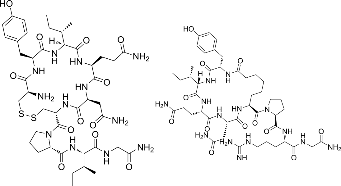
Fig. 2. The chemical structures mesotocin (left) and vasotocin (right) (National Center for Biotechnology Information, 2024).
This suggests a biochemical basis for social cohesion and flocking behavior. Ondrasek and her colleagues at UC Davis were the ones to come to this conclusion. They did so by using the Manning compound, which is known to have a higher attraction to vasopressin receptors than oxytocin receptors in mammalian brains. Thus, they predicted that the Manning compound would be more attracted to vasotocin receptors than mesotocin receptors in avian brains due to their structural similarities to vasopressin and oxytocin receptors, respectively. All subjects studied showed high levels of 125I-OVTA (125I-ornithine vasotocin analog) binding in the lateral septum, dorsal arcopallium, lateral arcopallium, and para-high vocal center, with the strongest signals occurring in the lateral septum (Table. 1 and Fig. 3). More moderate, 125I-OVTA binding occurred in other various portions of the nidopallium. 125I-LVA (125I-linearized vasopressin antagonist) binding almost completely overlapped with 125I-OVTA binding sites, except for the caudomedial mesopallium. The comprehensive map of these nonapeptide receptors resulting from this experiment now provides a basis for future studies on neuroecological reasons for collective behavior.
Table. 1. (left) Abbreviations for avian brain anatomical features. (right) Mean optical binding density of 125I-OVTA (125I-ornithine vasotocin analog) and 125I-LVA (125I-linearized vasopressin antagonist) in cerebral regions with visible binding in the European starling (Ondrasek et al., 2018).


Fig. 3. Representative photomicrographs of 125I-ornithine vasotocin analog (125I-OVTA; A, C, E, G, I, K, M, O) or 125I-linearized vasopressin antagonist (125I-LVA; B, D, F, H, J, L, N, P) binding in the brain of a European starling. Created via autoradiography assays (Ondrasek et al., 2018)
Olfactory system and social interactions
The existence of a functional olfactory system in birds has long been debated (Caro & Balthazart, 2010). Unlike many mammals that actively sample odors by extending their necks, moving their heads, and sniffing, birds typically do not exhibit such behaviors to assess the chemical composition of their environment. Due to the complexity of their multi-channel communication system involving acoustic and visual features, to be discussed shortly, birds were historically assumed to be poor at smelling. However, this is a misconception and birds are not anosmic.
The avian olfactory system shares many anatomical similarities with amphibians, mammals, and reptiles (Balthazart & Taziaux, 2009). The nasal cavity is fully developed with paired external nares that lead air into three internal chambers, with the third chamber containing olfactory receptors. These receptors detect chemical signals in the form of volatile organic compounds, which then connect to the brain via olfactory nerves and olfactory bulbs. Relatedly, research has proven evidence of neural projections from the olfactory bulbs to various brain regions, including the piriform cortex, mesopallium, and medial striatum, just like in mammals.
In starling flocks, the olfactory system aids in navigation and orientation, as demonstrated by studies showing that anosmic starlings—those with an impaired sense of smell—struggled to home over distances of 120 km or more (Wallraff et al., 1995). For example, only eight out of 101 groups of anosmic starlings were able to home from more than 100 km. This small success rate is attributed to one or many of several factors such as random search and chance, the possibility that the anosmic bird flew back with an olfactory bird, or that the ablation of the olfactory nerve was incomplete.
Recent studies suggest that species-specific odors are likely to play an important role in the control of social interactions in birds, such as mating and mother-offspring relationships (Balthazart & Taziaux, 2009). However, it is premature to claim that any avian odor signal functions as a pheromone (a species-specific chemical signal capable of eliciting a developmental or a physiological response). Extraneous odors have also been proven to interfere with the relationship between a youngling and its mother. These signals may trigger the parents’ abandonment and cessation of feeding of the younglings. Careful observations of starlings have demonstrated increased olfactory acuity as well as a beak color change during the breeding season. Enhanced olfactory acuity suggests that starlings can detect subtle chemical changes in the environment to aid in mate selection, such as the scent of competitors and the presence of other individuals in their territory. Furthermore, the chemical cues which birds interact with during the breeding season also reflect a change in sex hormones (Saavedra et al., 2022).
Gonadal development effect on flocking behavior
Flocking of starlings occurs in one of two distinct types (Davis, 1970). The first type, wherein starlings occur only in pairs, unfolds during the breeding season, approximately encompassing the months between February and July. The second type, which occurs during the non-breeding season of August through January, sees starlings form massive, coordinated flocks that number up to the millions. Additionally, in the breeding season, starlings tend to display heightened aggression towards members of their own species, often fiercely defending territories. This dramatic shift in behavior suggests a strong neurochemical influence (Davis, 1970).
Observations have suggested this is attributable to the level of sex hormones in the blood. Sex hormones, which is used rather broadly, encompasses testosterone in males, estradiol and other estrogens and progesterone in female starlings. Bullough concluded that as gonads decrease in size, reaching their smallest size in July and August, the amount of sex hormone released into the blood decreases, and birds are prompted to migrate southward in large flocks (Saavedra et al., 2022).
As one looks at a plot of average monthly flock size against gonadal volume (an indicator of the level of sex hormone produced) in male starlings, it is apparent that as gonadal size increases, average flock size decreases (Fig. 4) (Bullough, 1942).

Fig. 4. Average starling flock by monthly periods in relation to average monthly volume of starling testes (Bullough, 1942).
Crowding regulation
Starling colonies consist of dense aggregations of birds moving in coordination. Crowding is inevitable and is a legitimate source of stress within flocks. Corticosterone (CORT) is a steroid adrenal hormone responsible for stress, metabolism, violence and aggression in starlings and other animals (De Boer & Koolhaas, 2017). Crowding can affect starling behaviour as well as modify the levels of corticosterone in the bloodstream, depending on the starling’s social role. Corticosterone is synthesized by the adrenal glands of the kidney. When a bird experiences stress, the hypothalamus produces corticotrophin-releasing hormones, which trigger the release of adrenocorticotropic hormone by the pituitary gland. This then prompts the adrenal glands to secrete corticosterone. This hormone’s precursor is cholesterol, which undergoes multiple conversions to become corticosterone (Fig. 5). After secretion, corticosterone circulates in the animal’s bloodstream. Receptors in target cells’ cytoplasm bind this hormone, translocating to the nucleus to promote RNA transcription. The systemic release of corticosterone targets various organs and tissues such as cardiovascular, metabolic, neurological, and reproductive tissues. Corticosterone is associated with an increase in blood glucose levels and a suppression of non-essential functions to increase alertness (Kiilerich & Prunet, 2011).

Fig. 5. Conversion of cholesterol to corticosterone by the adrenal glands (Kiilerich & Prunet, 2011).
Heart rate is also affected by crowding. Crowding stimulates aggression within conspecifics, which may be a source of chronic stress that can be highly detrimental to individuals. This aggressive behavior can be regulated by hormones such as AVT, a peptide hormone known as arginine vasotocin. In birds, AVT can regulate blood pressure, and it was found to affect endocrine, behavioral and cardiovascular responses in other vertebrates. Additionally, behavior regulation through AVT could help improve starling fitness. Indeed, AVT was found to help significantly attenuate aggressive behavior between starlings by regulating stress responses through a cardiovascular mechanism, and not through the modification of CORT levels within the bloodstream (Fig. 6). Higher levels of AVT in the bloodstream decreased the frequency of maintenance behaviors of starlings such as drinking and feeding, as well as social aggressivity. This might be explained by the reduction of heart rate brought forth by surges in AVT, as a decreased heart rate leads to a slower metabolism, and hence, a slowing down of activity within starlings (Nephew et al., 2005).

Fig. 6. Starling activity and heart rate (BPM) following AVT injections of 1 or 4 μg or LRS (Ringer’s Lactate Solution as control) and immediate introduction to crowding. Activity and heart rate (HR) peak within minutes after injection (black arrow). 4µg AVT injection is associated with lower peaks in HR and activity than 1µg AVT the control. Mean HR returns to a stable state within fifteen minutes following injection, lower than baseline. Activity returns to baseline levels (Nephew et al., 2005).
Stress Response Mechanisms in Starlings
Vertebrate stress responses are vital for maintaining homeostasis and providing the body with proper defense mechanisms. A typical stress response (e.g. due to predator attack) occurs biphasically and mobilizes two distinct stress response systems within the body. The fight-or-flight response, initiated by the sympathetic nervous system, is induced due to imminent external stresses and provides the animal sufficient energy to escape a threat. The hyperactive sympathetic nervous system will stimulate the release of adrenaline, thereby triggering a physiological state of alertness which includes the increase of heart rate, blood pressure, and blood glucose levels (Awerman & Romero, 2010). The activation of the hypothalamic-pituitary-adrenal (HPA) neuroendocrine system also occurs as a response to stress. This system is a negative feedback mechanism which culminates in the release of glucocorticoids, which play an important role in glucose metabolism (Fig. 7). Indeed, the HPA axis inhibits hypothalamic function to prevent chronic hyperactivity of the sympathetic nervous system. In tandem with glucocorticoid-release, this is achieved by reducing corticotropin-releasing hormone (CRH) secretion by the hypothalamus. The fight-or-flight response will cease once CRH is no longer secreted (Gjerstad et al., 2018).

Fig. 7. Hypothalamic-pituitary-adrenal neuroendocrine system. This system is involved in the secretion of glucocorticoids (mainly corticosterone in birds) via an efferent regulatory pathway. CRF is a corticotropin-releasing factor and ACTH is an adrenocorticotropic hormone (Scanes, 2014).
The fight-or-flight response and the HPA axis both provide the system with additional energy by regulating glucose levels in the body. This is achieved either by appetite stimulation, glycogenolysis (hydrolysis of glycogen) or gluconeogenesis (synthesis of glucose) (Fig. 8). Other mechanisms of glucose regulation as a response to stress include triglyceride hydrolysis, the breakdown of muscle tissues, and the inhibition of glucose storage (glucocorticoids are insulin antagonists) (Awerman & Romero, 2010). Hydrolysis of triglycerides in adipocytes is also stimulated by glucocorticoids and the HPA axis, thus providing an efficient means for the body to maintain glucose levels as well as synthesize ATP for crucial high-energy responses (Baynes & Dominiczak, 2024).

Fig. 8. Glycogenolysis (bottom-to-top) and gluconeogenesis (top-to-bottom) pathways. Glycogen is a branched storage polysaccharide of glucose (Baynes & Dominiczak, 2024).
Chronic stress, HPA Suppression and Corticosterone Regulation
A chronically stressed system will have devastating impacts on the organism (Dickens et al., 2015). Chronic stress will affect reproductive and cellular function (e.g. affect immune function and somatic growth) and contribute to significant weight losses through wasting of the muscle mass. A study investigating the physiological effects of chronic stresses on starlings using blood biochemical parameters found that muscle degeneration is the main mode of disruption induced by chronic stresses (Awerman & Romero, 2010). This was achieved by conducting two succeeding stress-recovery cycles, wherein captive European starlings were subjected to multiple stressors each day during stress periods and deprived of it during recovery periods. Stressors included disturbances such as tapping their cage, subjecting them to loud noises and restraining them in confined of crowded spaces (Awerman & Romero, 2010). Capture myopathy, severe muscle damage accrued because of chronic overexertion due to stress, was identified as a major contributor to muscle degeneration along with fatty acid deficiency over time. Relatedly, corticosterone, the glucocorticoid secreted by the HPA axis in birds as a response to stress, will both inhibit protein synthesis and stimulate protein catabolism, thus further contributing to weight loss (Fig. 9). Interestingly, starlings that experienced significant weight loss during stress periods often gained more than their initial weight during recovery. This reveals innate adaptive measures in European starlings to prevent excessive damage due to stressors.

Fig. 9. Total protein, albumin, and globulin content in plasma after the initiation of Chronic Stress Period 1 (CSP1). Shaded regions indicate periods during which starlings were exposed daily to stressors. A decrease in albumin and globulin content, in this scenario, indicates metabolism of these proteins for energy. An increase in total protein plasma content during the second stress period indicates muscle degradation (Awerman & Romero, 2010).
Circulating glucocorticoids in the avian body affect more than merely protein metabolism. Their wide-ranging effects on reproduction and immunity make them a highly regulated chemical (Dickens et al., 2015). Whereas high reactivity to external stress as conferred by the HPA axis is beneficial in life-threatening conditions, prolonged overstimulation can lead to disease and malfunction. The fine balance between appropriate defense responses and chronic stress is programmed into birds through their breeding cycle. During the breeding season, when high system reactivity is of great importance for fitness and reproduction, the HPA axis in birds is highly active. Simultaneously, the greatest glucocorticoid release in response to stress occurs during the breeding season, indicating a more responsive system. On the other hand, during molt, when birds shed their feathers to assist new growth, the HPA axis observes a steep decline in activity, which is reflected by poorer glucocorticoid release in response to stress. It has been shown that both baseline and stress-induced glucocorticoid release may decline by up to 75% during the transition from breeding to molt (Dickens et al., 2015). Corticosterone, the primary glucocorticoid in birds, promotes HPA plasticity (Fig. 10). Therefore, higher levels of corticosterone in birds during breeding season would provide a more adaptive and dynamic HPA axis, which has been shown to increase individual fitness, and directly reproductive fitness. Contrastingly, corticosterone has also been shown to worsen the quality of growing feathers during molt. This exemplifies how lower levels of corticosterone during molt, and parallelly a less responsive HPA axis at that same time, would be beneficial for long-term fitness in birds (Dickens et al., 2015).

Fig. 10. Chemical structure of corticosterone. This steroid is secreted as a terminal hormone of the HPA axis and is involved in various physiological processes such as protein metabolism and reproduction. It also has stimulating effects on HPA plasticity and responsiveness to stress in an organism (Corticosterone (17-Deoxycortisol) | Endogenous Glucocorticoids | MedChemExpress, n.d.).
The growing of feathers is an energetically costly and complex process (Dickens et al., 2015). Much energy is dedicated to the growth of new feathers because their quality will affect critical aspects of a bird’s fitness such as flight, insulation and visual display. Corticosterone worsens feather quality in two ways. First, it affects the mechanism by which proteins are deposited into growing feathers, thus fundamentally altering protein assembly and structure. Second, high concentrations of corticosterone stimulate the bird and promote responsiveness, resulting in the expense of critical energy for other purposes than feather growing. The downregulated release of corticosterone during molt is an adaptation to maximize both the short-term and long-term survival of avian species (Dickens et al., 2015).
Chemistry of the visual system
Photoreceptors are specialized cells located at the back of the retina that absorb light and convert it into electrical signals through biological processes. Birds have two types of photoreceptors: rods and cones. Rods are responsible for vision in low-light or nighttime conditions (acuity). Cones are responsible for color vision and are more active during daylight (resolution). The distribution of rods and cones in a bird’s retina varies depending on its lifestyle. Nocturnal birds have a higher density of rods to maximize low-light vision. On the other hand, diurnal birds have more cones relative to rods, enhancing their ability to see fine details in bright light (Ruggeri, 2010). Birds possess one type of rod, one type of double cone, and four types of single cones. The four single cone types are sensitive to red, green, blue, and violet or ultraviolet light (Fig. 11).

Fig. 11. Photoreceptors in birds and their corresponding sensitivity to different wavelengths of light (Martin & Banks, 2023).
Photopigments are light-sensitive molecules found within photoreceptor cells that undergo chemical changes when they absorb light. Rhodopsin is found in rod cells. Rhodopsin is composed of opsin and 11-cis-retinal. (Fig 12). Rhodopsin only has one type of opsin that is highly sensitive to low light but not useful for distinguishing colors. In contrast, cone cells have four types of cone opsins each responsible for detecting specific wavelengths of light: red, green, blue, and violet or ultraviolet light. Each type of cone contains a specific opsin, thus allowing the cone to detect a particular range of wavelengths.
Opsins are proteins composed of about 350 amino acids with seven alpha helices embedded in the lamellar membrane of photoreceptor cells’ outer segments. The alpha-helices are primarily made up of non-polar amino acids, while the linkages between these helices contain polar amino acids. Opsin binds to a chromophore, specifically the retinal. The retinal undergoes isomerization from its 11-cis form to an all-trans configuration when it absorbs light. This change in the retinal induces a conformational change in the opsin, triggering a cascade of chemical reactions known as phototransduction. This process ultimately generates a nerve impulse that is transmitted to the brain (Hart, 2001).
The combination of opsin and retinal forms a visual pigment, with opsins determining the specific range of light wavelengths a cone is sensitive to through a process called spectral tuning. This phenomenon occurs when the polar amino acids near the retinal binding site in the opsin create electronic interactions with the retinal. These interactions change the amount of energy needed to excite the retinal from its ground state. The change in energy results in a change in the wavelengths of light that the visual pigment absorbs, which is known as an opsin shift. By altering the wavelengths absorbed by the visual pigment, the opsin shift directly affects the light wavelengths to which the cone is sensitive (Hart, 2001).
There are two types of opsin shifts: the bathochromic shift and the hypsochromic shift. A bathochromic shift occurs when the visual pigment absorbs light of lower energy and longer wavelengths than usual. This happens when opsin creates a binding pocket for the retinal that establishes a local electrostatic environment, which stabilizes the excited state of retinal more than the ground state. By stabilizing the excited state, the energy required for the retinal to reach this state is reduced (Hart, 2001).
On the other hand, a hypsochromic shift occurs when the opsin’s binding pocket creates a local electrostatic environment that stabilizes the ground state of the retinal more than the excited state. As a result, the visual pigment absorbs light of higher energy and shorter wavelengths. A hypsochromic shift can also occur if the opsin reduces electron delocalization in the retinal molecule. The 3D shape of opsin and the placement of nearby polar amino acids affect whether retinal absorbs longer wavelengths (bathochromic shift) by stabilizing its excited state, or shorter wavelengths (hypsochromic shift) by stabilizing its ground state. These molecular interactions explain why different opsins absorb distinct wavelengths of light. This molecular tuning acts as a design solution, enabling opsins to adjust their light absorption ranges and optimize the sensitivity of visual pigments across the whole spectrum. (Hart, 2001).

Fig. 13. The structure of a cone cell of bird (Jimfbleak, 2008).
In addition to opsin-retinal interactions, birds possess a unique feature called oil droplet in their cone cells, which are pigmented structures positioned in front of the opsins (Fig. 13). They are responsible for filtering the incoming light from the inner segment of photoreceptors before it reaches the visual pigment in the outer segment. For birds, oil droplets are often brightly colored due to the presence of carotenoid content. Carotenoids absorb shorter wavelengths of light, which allows only longer wavelengths to pass through. Therefore, oil droplets act as a lowpass “filter” which shifts the cone's sensitivity towards longer wavelengths than it would naturally detect. Additionally, oil droplets help narrow the range of wavelengths to which the cone is sensitive. This ensures that different types of cones have minimal overlap in their sensitivity. By reducing this overlap, oil droplets enhance the bird’s ability to discriminate between different colors. Together, the binding of retinal to opsin and the filtering effects of oil droplets play a crucial role in shaping the cone’s sensitivity and optimizing color vision in birds (Hart, 2001).
Energetics
Flight is the most energy intensive mechanical process carried out among all animals (Homan et al., 2011). Research aimed at analyzing the basal and active metabolic requirements of birds seeks to explain the mechanism by which birds can sustain high energy demands for long periods of time. Thus, as mentioned in our previous essay, morphological and physiological adaptations are primarily responsible for birds’ endurance during flight. Surprisingly, birds' metabolic machinery is largely unchanged relative to other non-flying animals. Here, we explore bird energetics from a chemical perspective (Homan et al., 2011).
Metabolic requirement model and meteorological effects
Metabolism refers to the ensemble of reactions involved in maintaining the basic function of any living organism. This includes breathing, blood flow, electric signals and maintaining the proper ions gradients. In essence, metabolism is the chemical process of maintaining homeostasis. The rate of metabolism varies considerably depending on the activity and the exterior conditions in which the organism finds itself. As a result, bird metabolic systems are subject to high levels of variation in demand. A study evaluating the validity of a bioenergetics model in caged European starlings (i.e. inactive) found that the basal true metabolic demand in birds varies between 208 kJ/bird and 274 kJ/bird every day (Homan et al., 2011). Their findings suggest that birds may consume anywhere between 157-305 kJ of food every day, although the true energy expenditure is generally lower due to heat generation during catabolism. To determine the relation between feeding rate and true energy loss during inactivity in European starlings, Equations 1 and 2 are employed:


Equation 1 describes the basal mass-dependent metabolic rate in homeotherms, where both C and e are constants specific to the species and m is the mass of the animal (kg). The e constant in passerines usually ranges between 0.6 and 0.7. Equation 2 describes the relationship between metabolic rate (M) and energy flux from the environment – λM denotes evaporative heat loss in the animal –. Relevant variables include air density ρ (kg/m3), the specific heat capacity of air cp (J/kgK), the animal’s body temperature Tb (K), the environmental temperature Te (K), the whole-body thermal resistance rb (W/K) and the sum of parallel resistances of the animal body’s surface comprising radiative heat loss and convective heat loss re (W/K). The latter resistances are added in parallel due to convection and radiation occurring simultaneously, as opposed to sequentially. These equations highlight two major characteristics of bird metabolism: they are dependent on mass and environmental conditions. The former constitutes significant proof as to why bird frames have decreased in size through evolution (see previous paper). Nonetheless, these equations allow bioenergetic modelling for the comparison of feeding rate and metabolic rate in starlings (Table. 3).
Table. 3. Table of feeding rate in starlings (kg/day) relative to meteorological variables. The true feeding rate M is compared with the daily metabolic rate as predicted by a bioenergetics model accounting for the effects of environmental conditions in energy losses. L95 and U95 are the lower and upper bounds for the confidence interval of the true feeding rate values, respectively (Homan et al., 2011).

Digestion itself contributes to energy expenditure as starling food intake levels are often higher than metabolic demand. Energy which does not contribute to active processes in the body is transduced into heat for thermoregulatory processes, which are of equal importance in maintaining homeostasis.
Glycolysis and cellular respiration
Starlings sustain their metabolic demands through their diet, which consists primarily of insects, including various crop pests (Rabie et al., 2023), and berries. In addition to this, starlings have been shown to consume nectar (Zungu & Downs, 2016). Catabolism refers to the process of breaking down larger or more complex molecules into smaller units to provide building blocks for an organism to synthesize biomolecules or to provide it with energy (Campbell & Reece, 2002). The latter is predominantly undertaken by glycolysis, the Krebs cycle, the electron transport chain and chemiosmosis, which consists in the oxidation and hydrolysis of glucose and its variants for the synthesis of adenosine triphosphate (ATP), commonly referred to as the energy currency in biological organisms. The sum of all these reactions, coined cellular respiration, grossly proceeds according to the following reaction:
C6H12O6 + 6O2 ⇒ 6CO2 + 6H2O + Energy
In addition to supplying the cell with ATP to sustain various metabolic processes, the above-mentioned reactions dissipate heat which enables homeothermic species to maintain their core body temperature. This heat which results from maintaining homeostasis is deemed obligatory thermogenesis (Silva, 2006). Thus, in colder environments, there are two hypothesized mechanisms by which organisms can sustain their body temperature. The first is by increasing the rate of ATP synthesis, which will generate more heat because of a greater number of chemical reactions in the cell. This can be achieved by the cell primarily through leakage ion channels. Indeed, the cell expends a significant amount of energy to maintain its homeostatic ion gradients (Silva, 2006). The second is by reducing the thermodynamic efficiency of the reaction system. This implies reducing the amount of work that can be done by the cell with a baseline number of chemical reactions (i.e. maintain reaction rate and synthesize less ATP). Poor thermodynamic efficiency in the electron transport chain occurs in the presence of uncoupling proteins. These proteins increase the permeability of the inner mitochondrial membrane to H+. If the membrane is permeable to H+, it cannot maintain an ion gradient, therefore no ADP can be phosphorylated by ATP synthase, which is responsible for ADP substrate phosphorylation. This renders the ETC reactions futile and increases the generation of heat by the cell (Silva, 2006).
Avian Longevity and arguments in favor of mitochondrial implication
There are many studied and confirmed correlations between the levels of reactive oxygen species (ROS) in living organisms and longevity. For instance, the mutation of mitochondrial polymerase gamma (Pol-G), which converts ROS into less reactive species, in mtDNA (mitochondrial DNA) mutator transgenic mice induced their premature ageing (i.e. alopecia, shortened lifespan, poor body condition) within six months. Relatedly, uncoupling proteins are also of particular interest in the study of longevity in avian species due to their effects on the mitochondria’s proton gradient (Fig. 14). Indeed, researchers posit that the impressive longevity of birds, both absolutely and with respect to mammals of similar sizes, might be attributable to their increased metabolic demands and body temperature, as well as respiration uncoupling (Hickey et al., 2012).

Fig. 14. Formation of a proton gradient across the inner mitochondrial membrane. This occurs because of the penultimate chemical process in the synthesis of ATP from glucose, the electron transport chain. NADH and FADH2, products of the Krebs cycle, are used as substrates in the ETC. Uncoupling proteins will render the membrane permeable to H+, thus uncoupling the ETC from the formation of a proton gradient (Clark et al., 2018).
Due to the requirement of flight, birds have high metabolic demands, resulting in aerobic frames and efficient fat-fuelled musculatures, which directly implies higher levels of fat in the muscles (Hickey et al., 2012). Fat can generate more ATP per mole than sugar, thus making it a valuable source of energy (Campbell & Reece, 2002). Additionally, birds maintain high core body temperatures of approximately 42°C, which is highest among vertebrates. While uncoupling proteins and proton leaks might contribute to maintaining the core body temperature of birds by making ATP synthesis less thermodynamically efficient, higher temperatures also result in increased membrane permeability, which would inherently increase respiration uncoupling (Fig. 14). Thus, to oppose positive uncoupling feedback, membrane composition is also regulated by interactions with proximal fatty acids to maintain a baseline permeability to maintain the proton gradient.
During oxidative phosphorylation, which combines the processes of the electron transport chain with chemiosmosis, high energy electrons leak from complexes I and III in the inner mitochondrial membrane to interact with molecular oxygen in the cell, thus forming ROS. Thus, mitochondria generate superoxide (O2-) as a byproduct of ATP synthesis via these complexes. Due to birds' aerobicity and high metabolic efficiency, it is hypothesized that their mitochondria will produce more heat at rest – less ATP, and by hypothesis, less ROS – due to respiration uncoupling. As the metabolic demand increases, during flight for instance, fat hydrolysis will recouple respiration, simultaneously providing birds with their required energy (Hickey et al., 2012).
It is also believed that mitochondria may serve as ROS sinks in the cellular environment (i.e. structures that deplete ROS levels in the cell). Indeed, several studies have shown a positive correlation between aerobic exercise and lifespan, wherein the effect is most remarkable in smaller organisms. Directly, aerobic exercise triggers mitochondrial biogenesis, thus explaining why mitochondrial mass relative to total mass is highest in birds and other active animals (Hickey et al., 2012). Respiring mitochondria dispose of hydrogen peroxide efficiently due to the enzyme system thioredoxin/peroxiredoxin which is coupled to the cycling process of NADP and NADPH in the cell. It is estimated that this enzyme system may remove up to 90% of hydrogen peroxide in the cell. Additionally, superoxide can reduce cytochrome c in the mitochondria, which can in turn reduce cytochrome c oxidase in the mitochondrial membrane, thus coupling superoxide oxidation to ATP synthesis (Hickey et al., 2012). In that case, oxidative stress would increase the rate of ATP synthesis in the cell. All indicated theories may contribute to increased longevity and lifespan in avian species due to the reduction of ROS by mitochondria.
Glucose Regulation and Adaptation
Birds maintain significantly higher glucose levels than other mammals. For instance, healthy avian blood glucose levels are approximately twice as large as those of like-sized mammals and between 200-400% higher than those of humans (Sweazea, 2022). This seemingly hyperglycemic state is hypothesized to be a consequence of the high metabolic demand of flight, although birds do not suffer from hyperglycemia-related pathologies. In the absence of metabolic stresses, birds maintain high glucose levels attributable to an acquired insulin resistance. This insulin resistance is believed to have originated during the Mesozoic era as a response to critically low atmospheric oxygen levels (Sweazea, 2022). These low-oxygen levels caused mass extinctions which drastically shrunk the theropod genome. This genome alteration enabled metabolic and anatomical modifications to occur. Although it was preserved in the liver, the insulin signalling pathway is believed to have been shut down in birds during this period, including the insulin receptor and insulin receptor substrate-1 (IRS-1), because of genetic losses. Chronically elevated levels of glucose and ketone bodies ensued, both of which are required substrates for ATP synthesis, which enabled birds to sustain high metabolic rates for intensive physical activity (Satoh, 2021).
The insulin adaptation is unique to birds and addresses their high metabolic demands in flight. Relatedly, when provided with low amounts of oxygen, and in the presence of insulin, there are two diverging mechanisms by which ATP synthesis can occur (Fig. 15).

Fig. 15. Two adaptations of ATP generation and glucose metabolism in low-oxygen environments. (A), adopted by most mammals, retains insulin-sensitivity to minimize ROS production by mitochondria. The end-product, lactate, results in the net phosphorylation of two ADP molecules. (B), adopted by birds, maximizes metabolism and energy generation in low-oxygen environments due to insulin-resistance. ROS leakage, which puts oxidative stress on the organism, is a consequence of this pathway (Satoh, 2021).
Mammals adopted a mechanism that reduces oxygen consumption, likely to minimize ROS production and oxidative stress (Satoh, 2021). Lactate fermentation, which results in metabolic rate suppression, allowed mammals to maintain insulin-sensitivity. Contrastingly, birds adapted insulin-resistance to maximize their metabolic rates despite low oxygen concentrations (Satoh, 2021). ROS leakage is a significant downside of this mechanism, although birds have constitutively activated their nrf2 gene as a resort. The nrf2 gene encodes nuclear factor erythroid 2-related factor 2, a protein which regulates antioxidant enzymes that boast protective capacities against oxidative damage (He et al., 2020).
Environmental effects
Effects on ecosystems
Starlings’ presence within local landscapes can greatly influence and shape ecosystems, another example of how colonial organisms can morph their surroundings. Intertwined in a complex food web, a singular disruption can trigger a chain of events.
The presence of starling colonies within rainforests of tropical Australia generates ecological hotspots, contributing to local biodiversity. Colonies composed of greater than a thousand starlings inhabit singular poison-dart trees (Antiaris toxicaria) between the months of November and April, where they nest and raise their offspring during the summer monsoon. At the end of the mating season, starlings migrate to New Guinea. They return to the same trees at each cycle, a routine which can last for upwards of 15 years. Nests crowd the branches, and aggregations of seeds and guano (bird feces high in nitrogen and phosphorus) accumulate beneath the tree, forming a “carpet” of nutrients (Natusch et al., 2016). Starlings are known to affect the quantities of nitrogen and phosphorus in their local environments, and areas with dense populations of birds could experience a heightened deposit of these elements. Thus, starlings could play a great role within their ecosystem by promoting nutrient cycling (Hayes et al., 1984).
During mating season, eggs and chicks often slip from the nests and gather on the forest floor. These attract pythons, dingoes as well as birds of prey which feed on them. It was found that, especially during mating season (wet season), soil invertebrates and flying invertebrates thrive more so beneath colony trees than trees uninhabited by starlings. Nutrients derived from starlings’ guano generate a favorable environment for soil invertebrates to proliferate. Twenty-five species of animals were detected under colony trees, and significantly more mammals as well as birds congregate beneath colony trees as opposed to regular trees (Natusch et al., 2016). Toads and snakes are abundant at night, and during the day, pigs often feed on roots and seedlings nourished by the rich soil churned by invertebrates. Vegetation proliferates, attracting herbivores and insectivores. This phenomenon is exacerbated during the dry season as resources within the forest dwindle, making these ecological hotspots even more important for species survival (Fig. 16). This explains why pigs, along with birds such as cockatoos, owls and turkeys tend to feed off residual nutrients left over after the mating season, during dry season. During mating season, predators tend to congregate beneath colony trees, which could also dispel these prey organisms from visiting those zones during the wet season.
Some species’ density under trees inhabited by colonies of starlings are up to a thousand times greater than anywhere else. In essence, some organisms benefit from the primary resources produced by starlings such as fallen chicks and eggs, whereas others derive benefit by consuming secondary resources produced by starlings such as roots and invertebrates. This demonstrates the complexity of food webs and how all resources are to be utilized in nature. The concentration of these nutrient-dense resources contributes to the aggregation of many species, creating heterogeneous zones of species richness within the wild. These dense zones fluctuate temporally and spatially depending on the climate, season and resource availability. Survival of species as well as rates of reproduction are affected by these zones, which suggests that there are fitness benefits tied to using these ecological hotspots (Natusch et al., 2016). Studying starlings’ effects on ecosystems can allow for the development of more effective conservation methods, potentially ensuring the survival of many wildlife species.

Fig. 16. Animal aggregations beneath starling-inhabited tree. a) Colony tree; b) Scrub pythons feeding on fallen chicks; c) Brush turkeys; d) Wild pigs foraging under a colony tree (Natusch et al., 2016).
Pathogenic effects
Starlings are often regarded as pests, as they decimate crop yields and exacerbate noise pollution. Since they are associated with human development and agriculture, there have been many concerns regarding their ability to act as disease vectors. Concentrated animal feeding operations (CAFO's) have exacerbated the development of antibiotic-resistant strains of bacteria as it allows for the rapid proliferation and mutation of microorganisms within cattle. CAFO’s can be sources of great microbial pollution to the environment (Carlson, 2011).
Around 50% of starlings are potential carriers of the bacterium Escherichia coli, and half of these individuals might carry a pathogenic strain (Cabe, 2021). It was found that the transmission of pathogenic E. coli to livestock seems to be enacted through contamination of water, as well as through the contamination of cattle feed due to starling guano. Certain pathogenic strains of E. coli can be transmitted from starling to cattle, and from cattle to humans, leading to public health concerns. A rather alarming phenomenon is the transmission of multiple antibiotic-resistant strains of bacteria to cattle which is documented circumstantially. Numerous genes control antibiotic resistance and could be spread from one bacterium to another through the integration of plasmids, or other mechanisms such as drug inactivation through the expression of enzymes which inactivate antibiotics. These strains were found to be resistant to a multitude of antibiotics such as ampicillin, streptomycin and β-lactam antibiotics, among many others.
Starlings are also proponents of salmonella infections among livestock and potentially humans. Guano can contain this pathogenic bacterium, and similarly to the transmission of E. coli, starling droppings can contaminate sources of water along with livestock food, passing on the disease to mammals. It was found that winter months may be more highly correlated with salmonella outbreaks within farms as starlings congregate and roost in large numbers (Cabe, 2021). The instauration of measures to counter the settling of starlings within CAFO’s is highly correlated with the reduction of S. enterica pathogens. Chemicals such as 3-chloro-4-methylaniline (Fig. 17), known as DRC-1339, are used as an effective way to control starling population, as it is a slow-acting starlicide (Carlson, 2011). Starlings and blackbirds are more sensitive to starlicide, metabolizing it rapidly. Irreversible heart and kidney damage follow due to impeded metabolic and cellular functions, resulting in renal failure and death 1-3 days after exposure. Other species are less sensitive but can experience nervous system depression along with respiratory or cardiac arrest (U.S. Department of Agriculture: Animal and Plant Health Inspection Service, 2001). The exact biochemical mechanism of starlicide has not been identified in the available literature, which instead focuses on its toxicological effects.

Fig. 17. 3-Chloro-4-methylaniline, also known as Starlicide, or DRC-1339, a lethal toxin for starlings (Serfas, 2009).
Numerous other bacterial infections among cattle can be attributed to the presence of starlings. For instance, Gastroenteritis Virus, a viral infection transmissible from starlings to pigs, results in incessant diarrhea and high mortality rates. Starlings could act as a vector for this pathogen, spreading it from one farm to another. The spread of fungal pathogens is an additional risk brought forward by starling colonies, as the ascomycete Histoplasma capsulatum can be contained within starling feces. The inhalation of these mold spores could lead to the development of pulmonary histoplasmosis within the human host, a lung disease which can be debilitating in certain cases (Wheat et al., 2009).
It should be noted that starlings’ presence can greatly benefit natural ecosystems through their droppings. In contrast, this same phenomenon has been found to disrupt human-dominated environments, bringing forth infection and death. It can be hypothesized that this dissonance is due to wildlife’s resilience to certain pathogens widespread in nature, as opposed to domestic animals and humans. Carriers must develop tolerance to survive. Due to constant exposure, birds’ immune systems have evolved to tolerate a certain microbial load without affecting their own survival. Additionally, in CAFO’s, restricted air flow and consistent conditions favor the spread of pathogens, whereas in nature, bird feces can be rapidly degraded by rain and microbes, leading to less accumulation of pathogens. Humans and farm animals have been subjected to more sterile environments than wildlife. Their immune systems have progressively lost the ability to handle such high microbial loads (Baucom & Roode, 2011).
Guano
Guano are mineralized deposits formed by the feces, eggshells, and carcasses of birds or bats. It has historically been used as an effective plant fertilizer due to its high phosphorus and nitrogen content, as its composition is beneficial for plant growth. Indeed, it also contains minimal amounts of zinc, copper, magnesium, chloride, calcium, sulfur, potassium, iron, and manganese, all proponents of plant growth (Schnug et al., 2018). Guano is mineralized under hot and dry climates, usually in subtropical coastal deserts under high pressure zones, typically around latitudes of 30°N and 30°S. Seabirds are the main producers of guano. Although starlings are not seabirds, their excrements also contain similar minerals which could aid plant growth, as seen beneath colony trees in Australian tropical forests. Guano can be characterized by its formation and age. White guano is the result of recent excretion. Produced continually by birds, it usually consists of 3% K2O, around 12% nitrogen, and 12% phosphoric acid (P2O5), a source of phosphorus. Red guano, on the other hand, is fossilized from geological occurrences, and can contain around 30% phosphoric acid, contributing high levels of phosphate within soils to which it is added (Schnug et al., 2018).
Guano can be classified as either nitrogen guano or phosphate guano. Phosphate guano is produced from the leaching out of soluble phosphate and nitrogen attributable to continuous interactions with seawater and rain. If this process takes place over formations abundant in calcium carbonate, rock guano can form. Rock guanos are rich in apatite—a compound made of calcium and phosphate—and have low nitrogen levels. Nitrogen-rich guanos, on the other hand, contain relatively more phosphate compared to nitrogen. However, their nitrogen levels are higher than phosphate guanos. Seabirds directly affect the composition of guano as the byproducts of their metabolism determine its composition. Higher levels of phosphate and higher protein intake in birds could affect the abundance of nitrogen and phosphate in guano. Relatedly, the end products of bird metabolism, such as urea, contain lots of ammonium, and hence nitrogen. Ammonium oxalate and ammonium urea are both derived from the metabolic processes of birds. Additionally, guanine bases in DNA might also contribute to guano’s nitrogen content (Schnug et al., 2018). Diet is quite important in determining phosphate concentration in guano, as a diet rich in fish leads to the production of guano rich in phosphate, and a diet rich in plankton results in guano with poorer phosphate content (Zwolicki, 2013). Although seabirds are most responsible for mineralized guano in coastal areas, starlings, as colonial animals, produce excrements in large quantities, which can nourish certain microenvironments. Starling guano can provide nutrients to plants in certain contexts (de Boer and Koolhaas 2017), however its hypothetical usage as commercial fertilizer could be disastrous due to its high pathogenicity (Carlson, 2011).
Conclusion
Metabolic regulation enables unique avian behaviours. Indeed, regulation is modulated to sustain a chronically hyperactive system while preventing overexertion and long-term physiological imbalances. This is critical because birds’ locomotion is reliant on the efficient expenditure of energy while their fitness is largely dependent on the structural quality of their physiological attributes. High metabolic rates in birds are maintained through a unique ETC uncoupling process. ETC uncoupling is preserved during metabolic downtime to increase body temperature. As metabolic demand increases, fat hydrolysis recouples the respiration process to generate more ATP. Metabolic and stress regulation are also coupled to birds’ mating cycles. The HPA axis, which promotes corticosterone release and plasticity in the sympathetic nervous system, is heavily downregulated during molt to improve feather quality, which exemplifies how short-term drawbacks in birds’ ability to generate a stress response can preserve their long-term fitness. Physiological adaptations in murmuring birds can also contribute to the murmuration’s improved cohesion and coordination. Oil droplets in birds’ eyes ensure that the wavelengths to which different opsins are sensitive are sufficiently distinguishable to maximize the resolution of their sight. Improving features essential for coordination during predatorial defense promotes the long-term survival of the species.
Studying the chemical adaptations of birds provides a unique opportunity to translate their ingenious mechanisms of survival to the benefit of all. Unique characteristics of birds, such as their refined sensitivity to ambient chemicals for coordination, or the factors which render their metabolism particularly efficient for long-term periods of exertion and longevity, offer academics and engineers alike a prominent model for innovation in the health sectors. Other relevant fields of interest in relation to avian life and physiology, such as their ecology or the agricultural and chemical value of their excreted matter, lay out undeniable foundations for establishing renewable and sustainable practices both in industry and in our interactions with other living organisms.
References
References
Awerman, J. L., & Romero, L. M. (2010). Chronic psychological stress alters body weight and blood chemistry in European starlings (Sturnus vulgaris). Comparative Biochemistry and Physiology, Part A. 10.1016/j.cbpa.2010.01.010
Balthazart, J., & Taziaux, M. (2009). The underestimated role of olfaction in avian reproduction?. Behavioural brain research, 200(2), 248–259. https://doi.org/10.1016/j.bbr.2008.08.036
Baynes PhD, J. W., & Dominiczak, M. H. (2024). Biomedical Chemistry (J. W. Baynes PhD & M. H. Dominiczak, Eds.). Elsevier.
Baucom, R. S., & de Roode, J. C. (2011). Ecological immunology and tolerance in plants and animals. Functional Ecology, 25(1), 1-10. https://doi.org/10.1111/j.1365-2435.2010.01742.x
Cabe P. R. (2021). European Starlings (Sturnus vulgaris) as Vectors and Reservoirs of Pathogens Affecting Humans and Domestic Livestock. Animals : an open access journal from MDPI, 11(2), 466. https://doi.org/10.3390/ani11020466
Campbell, N. A., & Reece, J. B. (2002). Biology. Benjamin Cummings.
Carlson, J.C., Engeman, R.M., Hyatt, D.R. et al. (2011). Efficacy of European starling control to reduce Salmonella enterica contamination in a concentrated animal feeding operation in the Texas panhandle. BMC Vet Res 7, 9. https://doi.org/10.1186/1746-6148-7-9
Caro, S. P., & Balthazart, J. (2010). Pheromones in birds: myth or reality?. Journal of comparative physiology. A, Neuroethology, sensory, neural, and behavioral physiology, 196(10), 751–766. https://doi.org/10.1007/s00359-010-0534-4
Clark, M. A., Choi, J. H., & Douglas, M. (2018). Biology 2e. OpenStax.
Corticosterone (17-Deoxycortisol) | Endogenous Glucocorticoids | MedChemExpress. (n.d.). MedchemExpress.com. Retrieved October 19, 2024, from https://www.medchemexpress.com/Corticosterone.html
Davis, G. J. (1970). Seasonal Changes in Flocking Behavior of Starlings as Correlated with Gonadal Development. Wilson Bulletin, 82(4), 391-399.
De Boer, S. F. and J. M. Koolhaas (2017). The Neurobiology of Offensive Aggression☆. Encyclopedia of Behavioral Neuroscience, 2nd edition (Second Edition). S. Della Sala. Oxford, Elsevier: 191-201. https://doi.org/10.1016/B978-0-12-809324-5.00382-5
Dickens, M. J., Vecchiarelli, H. A., Hill, M. N., & Bentley, G. E. (2015). Endocannabinoid Signaling in the Stress Response of Male and Female Songbirds. Endocrinology. https://doi.org/10.1210/en.2015-1425
Gjerstad, J. K., Lightman, S. L., & Spiga, F. (2018). Role of glucocorticoid negative feedback in the regulation of HPA axis pulsatility. The International Journal on the Biology of Stress.
https://doi.org/10.1080/10253890.2018.1470238
Hart, N. S. (2001). The Visual Ecology of Avian Photoreceptors. In Progress in Retinal and Eye Research (Vol. 20, Issue 5, pp. 675–703). Elsevier BV. https://doi.org/10.1016/s1350-9462(01)00009-x
He, F., Ru, X., & Wen, T. (2020). NRF2, a Transcription Factor for Stress Response and Beyond. International Journal of Molecular Sciences. 10.3390/ijms21134777
Hickey, A., Jüllig, M., Aitken, J., Loomes, K., Hauber, M., & Phillips, A. (2012). Birds and longevity: Does flight driven aerobicity provide an oxidative sink? Ageing Research Reviews, 11.
https://doi.org/10.1016/j.arr.2011.12.002
Homan, J., Stahl, R., & Linz, G. (2011). Comparing a Bioenergetics Model With Feeding Rates of Caged European Starlings. Journal of Wildlife Management. 10.1002/jwmg.19
Jimfbleak. (2008). Bird cone cell, including oil droplet [Digital image]. Wikimedia Commons. https://en.wikipedia.org/wiki/Oil_droplet#/media/File:BirdCone.png

Kiilerich, P., & Prunet, P. (2011). Hormonal control of metabolism and ionic regulation: Corticosteroids. In A. P. Farrell (Ed.), Encyclopedia of fish physiology (pp. 1474–1482). Academic Press. https://doi.org/10.1016/B978-0-12-374553-8.00186-6
Martin, G. R., & Banks, A. N. (2023). Marine birds: Vision-based wind turbine collision mitigation. In Global Ecology and Conservation (Vol. 42, p. e02386). Elsevier BV. https://doi.org/10.1016/j.gecco.2023.e02386
National Center for Biotechnology Information (2024). PubChem Compound Summary for CID 68649, [Arg8]vasotocin. Retrieved November 12, 2024 from https://pubchem.ncbi.nlm.nih.gov/compound/Arg8_vasotocin.
National Center for Biotechnology Information (2024). PubChem Compound Summary for CID 92135813, Mesotocin. Retrieved November 12, 2024 from https://pubchem.ncbi.nlm.nih.gov/compound/Mesotocin.
Natusch, D. J., Lyons, J. A., Brown, G., & Shine, R. (2016). Communally Nesting Migratory Birds Create Ecological Hot-Spots in Tropical Australia. PloS one, 11(10), e0162651. https://doi.org/10.1371/journal.pone.0162651
Nephew, B. C., Aaron, R. S., & Romero, L. M. (2005). Effects of arginine vasotocin (AVT) on the behavioral, cardiovascular, and corticosterone responses of starlings (Sturnus vulgaris) to crowding. Hormones and Behavior, 47(3), 280-289. https://doi.org/10.1016/j.yhbeh.2004.11.007
Ondrasek, N. R., Freeman, S. M., Bales, K. L., & Calisi, R. M. (2018). Nonapeptide Receptor Distributions in Promising Avian Models for the Neuroecology of Flocking. Frontiers in Neuroscience, 12. 10.3389/fnins.2018.00713
Rabie, D., Farhi, K., & Chedad, A. (2023). What does the diet of the Common Starling (Sturnus vulgaris Linnaeus, 1758) in the Ziban Oasis (Algeria) indicate: pest or useful in agriculture? Zoology and Ecology, 33. 10.35513/21658005.2023.2.10
Ruggeri, M., Major, J. C., Jr, McKeown, C., Knighton, R. W., Puliafito, C. A., & Jiao, S. (2010). Retinal Structure of Birds of Prey Revealed by Ultra-High Resolution Spectral-Domain Optical Coherence Tomography. In Investigative Opthalmology & Visual Science (Vol. 51, Issue 11, p. 5789). Association for Research in Vision and Ophthalmology (ARVO). https://doi.org/10.1167/iovs.10-5633
Saavedra I, Tomás G, Amo L. Assessing behavioral sex differences to chemical cues of predation risk while provisioning nestlings in a hole-nesting bird. PLoS One. 2022 May 19;17(5):e0268678. doi: 10.1371/journal.pone.0268678. PMID: 35588122; PMCID: PMC9119470.
Satoh, T. (2021). Bird evolution by insulin resistance. Trends in Endocrinology and Metabolism.
https://doi.org/10.1016/j.tem.2021.07.007
Scanes, C. G. (Ed.). (2014). Sturkie's Avian Physiology. Elsevier Science.
Schnug, E., Jacobs, F., & Stöven, K. (2018). Guano: The White Gold of the Seabirds. InTech. doi: 10.5772/intechopen.79501
Serfas et al. (2009). Starlicide. Wikipedia, https://en.wikipedia.org/wiki/Starlicide
Silva, E. (2006). Thermogenic Mechanisms and Their Hormonal Regulation. Physiological Reviews, 86. 10.1152/physrev.00009.2005
Sweazea, K. L. (2022). Revisiting glucose regulation in birds – A negative model of diabetes
complications. Comparative Biochemistry and Physiology Part B: Biochemistry and Molecular Biology. https://doi.org/10.1016/j.cbpb.2022.110778
U.S. Department of Agriculture: Animal and Plant Health Inspection Service. (2001, April). DRC-1339 (Starlicide). DigitalCommons@University of Nebraska - Lincoln. https://digitalcommons.unl.edu/cgi/viewcontent.cgi?article=1735&context…
Wallraff, H. G., Kiepenheuer, J., Neumann, M. F., & Streng, A. (1995). Homing Experiments with Starlings Deprived of the Sense of Smell. The Condor, 97(1), 20-26. https://doi.org/10.2307/1368979
Wheat, L.J., Connolly, P., Smedema, M., Rogers, P.D. (2009). Antifungal Drug Resistance in Histoplasmosis. In: Mayers, D.L. (eds) Antimicrobial Drug Resistance. Infectious Disease. Humana Press. https://doi.org/10.1007/978-1-60327-595-8_21
Zungu, M., & Downs, C. (2016). Digestive efficiencies of Cape white-eyes (Zosterops virens), red- winged starlings (Onychognathus morio) and speckled mousebirds (Colius striatus) fed varying concentrations of equicaloric glucose or sucrose artificial fruit diets. Comparative Biochemistry and Physiology, Part A, 199. 10.1016/j.cbpa.2016.05.004
Zwolicki, A., Zmudczyńska-Skarbek, K.M., Iliszko, L. et al. (2013). Guano deposition and nutrient enrichment in the vicinity of planktivorous and piscivorous seabird colonies in Spitsbergen. Polar Biol 36, 363–372. https://doi.org/10.1007/s00300-012-1265-5