


 The GCI's Histology Facility provides a full range of histology services to the McGill research community as well as the surrounding academic and private sectors at very competitive prices.
Under the guidance of a board certified veterinary comparative pathologist, our highly trained histology technicians provide the highest standards in histotechnology services. Under strict quality control surveillance, your samples will be processed in quick turn-around time. Your satisfaction is our pride.
Contact us:
Office 514-398-5647
Lab 514-398-8270
Email histology.gcrc [at] mcgill.ca
Contact names:
Plinio Da Cruz (plinio.queirozdacruz [at] mcgill.ca)
Nicole Robinson (nicole.robinson2 [at] mcgill.ca)
Grossing, often referred to as “cut-up”, involves a careful examination of the specimen to ensure that the proper area is chosen and the specimen is the proper thickness. Larger specimens may require further dissection to produce representative pieces from appropriate areas. For example multiple samples may be taken from the excision margins of a tumor to ensure that the tumor specimen has normal tissue surrounding it to check for infiltration. In the case of small specimens the entire specimen may be processed. The tissues selected for processing will be placed in cassettes (small perforated baskets) and batches will be loaded onto a tissue processor for processing through to wax.
If you are not sure what or how you should be submitting your samples we can assist you.
Processing: Tissue Tek VIP5 Vacuum Infiltration Processor
The GCRC Histology Core Facility uses an automatic, self-contained tissue processor, which holds up to 300 cassettes. The VIP software is programmable for up to 20 different programs for use in the fixation, dehydration, clearing and paraffin of a variety of specimens. We can customize programs to the specific needs of our clients.
Embedding: Leica EG 1150H
The Leica EG1150 modular tissue embedding center incorporates two separate components, the independent modules offer the flexibility to arrange embedding workflow for maximum efficiency.
Tissue is oriented in a metal mold filled with hot paraffin.
Cutting
Equipped with various automatic and manual microtomes our staff will provide publication quality paraffin sections from 4 microns or thicker.
There is 1 Leica microtome dedicated for student use.
Staining
The Leica ST5020 Multistainer produces consistent, high-quality results for both routine and special stains and can perform single or multiple protocols at the same time.
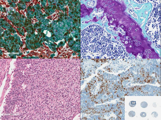
Special Stains
We have a large choice of special stains to choose from.
Can’t find the stain you need? Contact us at histology.gcrc [at] mcgill.ca for other possibilities.
| Stain | Results | Stain | Stain |
|
Congo Red |
Amyloid |
Oil Red O |
Lipids |
|
Cresyl Violet |
Nissel Substances |
P.A.S |
Glycogen,mucin & some basement membrane |
| Verhoff |
Elastic Fibers |
P.A.S./Alcian Blue |
To demonstrate the full complement of tissue proteoglycans |
|
Wright Geimsa |
Facilitates the differentiation of blood cell types |
Prussian Blue |
Iron |
|
Gomory Reticulin |
Reticulum fibers |
Toluidine Blue |
Mast Cells |
| Gram | Bacteria |
Von Kossa |
Minerals |
|
Grocott (GMS) |
Fungi |
Warthin-Starry |
Microorganisms |
|
Luxol Fast Blue |
Mylene |
Ziehl-Neelsen |
Acid fast Bacilli |
|
Masson’s Trichrome |
Collagen | ||
| Mucicarmine | Mucin | ||
|
Nuclear Fast Red |
Nuclear |
The histology core is excited to announce a new service for automated immunohistochemistry.
We have established protocols for the following markers:
Other markers may be developed upon request, as long as an appropriate antibody is available.
Validated IHC Stains
| Marker |
Target Population |
Target species |
Notes |
|
ATP6IP2 (aka PRR) |
Cells expressing ATP6IP2 |
Mouse, human |
Specificity of staining uncertain |
|
Beta-catenin |
cells expressing beta-catenin |
Mouse, rat, human, primate |
|
|
CEACAM-1 |
cells expressing CEACAM-1 |
Mouse, human |
Mouse monoclonal antibody |
| CD3 |
T lymphocytes |
Mouse, human, rat |
|
| CD31 |
Endothelial cells |
Mouse only |
|
| CD31 |
Endothelial cells |
Rat only |
|
|
CD34 |
Mesenchymal and hematopoietic stem cells, endothelium |
Human | |
| CD34 |
Mesenchymal and hematopoietic stem cells, endothelium |
Mouse, rat, others |
|
| CD79a |
B lymphocytes |
all except mouse |
|
|
Cyclin D1 |
cells in proliferative phase |
mouse, human, rat |
|
|
CC3 (cleaved caspase-3) |
Apoptotic cells |
Mouse, human, rat |
|
|
GFAP (glial fibrillary acidic protein) |
Astrocytes |
All | |
|
phospho-Histone H2A.X |
Cells expressing phosphorylation of Histone 2A.X at ser139 (indicating DNA damage) |
Mouse, human, rat |
May also stain cells undergoing division |
|
phospho-Histone H3 |
Cells in mitotic phase |
Mouse, human |
|
|
Iba-1 |
Macrophages |
Mouse, human, rat, primate |
|
|
Ki-67 |
Proliferating cells (all cell cycle phases) |
Mouse, human, others |
|
|
Melanoma gp100 |
Melanoma cells |
Mouse, human |
used with red chromogen (AEC) to distinguish from melanin |
|
MBP (myelin basic protein) |
Myelin (cerebral white matter, nerves) |
All | |
|
NoGo-A |
Oligodendrocytes |
Mouse, rat, human, primate |
Cytoplasmic marker |
|
Olig-2 |
Oligodendrocytes |
Mouse, rat, human, primate |
Nuclear marker |
| p53 |
Cells overexpressing p53 |
Mouse, rat |
Should recognize wild and mutant p53 |
|
pan-cytokeratin (AE1/AE3) |
Most epithelia (cytokeratins 1,2,3,4,5,6,7,10,14,15,16,19) |
all except mouse |
Does not react with cytokeratin 18 (most hepatocellular carcinomas negative). May react with glial cells and tumors |
| Parvalbumin |
Parvalbumin-positive neurons |
Human | |
|
phospho-SMAD3 (ser423/425) |
Cells expressing SMAD3 phosphorylation at ser423/425 |
Mouse, human, primate |
|
|
phospho-STAT (tyr405) |
Cells expressing STAT3 phosphorylation at tyr705 |
Mouse, human, primate |
Staining highly dependent on tissue fixation conditions |
| Somatostatin |
Cells producing somatostatin |
Human | |
|
Spondin 2 |
Cells expressing spondin 2 |
Mouse, human |
Specificity of staining uncertain |
|
UCP-1 |
Cells expressing UCP-1 |
Mouse, rat, others |
Specificity of staining uncertain |
|
vWF (vonWillebrand factor) |
endothelial cells, platelets |
Rat | |
| In preparation VEGF |
Our qualified histology technicians can provide training to staff and students in:
The histology core provides a dedicated student room equipped with a Fisher embedder, a Leica microtome, and 2 cryostats. (Microm & Fisher Cryotome). The student room is available 24/7 all year round.
TMA Grand Master (currently the only one in Canada)
Highest capacity: 72 blocks
• 60 donor blocks at the same time
• 12 recipient blocks
4 core diameters
• 0.6 mm max. 558 cores
• 1 mm max. 286 cores
• 1.5 mm max. 135 cores
• 2 mm max. 84 cores
Fastest microarrayer
• 2 mm max. 84 cores
• max. 12 seconds per core
• Simultaneous loading, imaging, drilling and punching
Smart automation
• Automatic block height measurement to ensure the embedded cores are in alignment with the recipient block surface
• Automatic barcode reading , donor block and label image saving for reference, project data saving into Excel file and Automatic PCR extraction
PCR extraction function
• 6 PCR cassettes
•10 PCR tubes/cassettes
Use of cleaning block to avoid cross contamination
Extracted FFPE tissue samples are ready for DNA extraction and PCR analysis with commercially available kits