The labs of Dr John White and Dr Jorg Fritz are seeking for an highly motivated postdoctoral fellow, who can begin in May or June 2018. The postdoc should have expertise in flow cytometry and mouse work, and competence in Bioinformatics would be an asset. The successful candidate will work in both lab environments.
Background.Vitamin D was first identified as a cure for nutritional rickets, a disease of bone growth caused by an inadequate uptake of dietary calcium. Cod liver oil was discovered as an excellent source of anti-rachitic activity in 1827, although it wasn’t until several decades later that the active ingredient was identified as vitamin D3. Even earlier, in 1822, a Polish physician experimenting with children reached the remarkable conclusion that sunlight cured rickets. It is now known that secosteroidal vitamin D3 is produced in skin via photochemical and thermal conversion of 7-dehydrocholesterol in the presence of UVB light. UVB irradiation is absorbed by atmospheric ozone; consequently, surface UVB varies markedly in intensity with latitude and time of year. At the latitude of Montreal UVB intensity is sufficient to make vitamin D in skin during 6 months of the year. Moreover, as vitamin D intake is generally inadequate in most diets, vitamin D insufficiency or deficiency rises in frequency with increasing latitude. Epidemiological studies link vitamin D deficiency to increased rates of cancer, as well as immune and infectious diseases. U.S. rates of bladder, breast, colon, ovary and rectal cancer increase 2-fold from south to north and north-south gradients of immune conditions such as multiple sclerosis, type 1 diabetes and Crohn’s disease have been documented. |
 COPYRIGHT 2007 SCIENTIFIC AMERICAN, INC. |
|
Much of the action of 1,25D can be explained by its binding to and activation of the vitamin D receptor (VDR). The VDR is a ligand-activated transcription factor composed of a highly conserved DNA binding domain, and an α-helical ligand binding domain. To find out more about vitamin D and the VDR, read our article in Scientific American [view PDF format]. |
|
Project 1(i). Regulation of antimicrobial innate immunity by vitamin D.
Given that the VDR is a transcription factor and acts as a ligand-regulated gene switch, its signaling is ideally suited to analysis using genomic approaches. We have used a combination of microarrays and in silico screens for VDREs to identify several hundred 1,25D target genes (Mol. Endocrinol. 2001 [view PDF format]; Mol. Endocrinol. 2002 [view PDF format]; Mol. Endocrinol. 2005 [view PDF format], EMBO Rep. 2006 [view PDF format] and have identified ~1000 novel target genes of 1,25D in squamous carcinoma cells. These studies led to the discovery that expression of gene encoding antimicrobial peptides (AMPs) CAMP (cathelicidin antimicrobial peptide, hCAP18, LL37) and DEFB2 (DEFB4, β-defensin 2) was under control of the vitamin D receptor (J. Immunol. 2004 [view PDF format]), demonstrating for the first time that vitamin D is a direct inducer of antimicrobial innate immunity in humans. Since then we have found that 1,25D regulates other genes that control innate immune responses, and are actively studying the mechanisms and physiological consequences of their regulation.
Vitamin D and Tuberculosis. Our current efforts are focused on elucidating the role of vitamin D signaling in combatting M. tuberculosisinfection of macrophages, the primary targets of M.tb. infection. We have performed extensive gene expression profiling studies and have found that vitamin D signaling has pleiotropic effect on infected macrophages, including enhancing the extensive cytokine/chemokine response elicited by infection (PLoS Pathogens, 2013 [view PDF format]). We are complementing gene expression profiling studies with genome-wide ChIPseq analyses to determine the host macrophage transcriptional response to infection.
Project 1(ii). Investigating the anticancer properties of vitamin D. |
|
|
Identifying 1,25D target genes underlying its anticancer effects. For the last several years, we have been analyzing the potential of analogs of vitamin D3 as potential agents of cancer chemoprevention. We have found treatment of squamous carcinoma cells with a vitamin D analog induces expression of several markers of squamous cell differentiation, and suppresses markers of squamous carcinoma progression in vitro and in vivo. (J. Nat. Cancer Inst. 2001 [view PDF format]). We have been focusing on the molecular events underlying cell cycle arrest by vitamin D signaling and have found that the VDR controls cell cycle progression in part by inducing the function of FoxO transcription factors, cell cycle inhibitors and bona fide tumor suppressors (Mol. Cell Biol., 2010 [view PDF format]). More recently we showed that vitamin D signaling through the VDR controls control expression and function of cMYC, an oncogenic transcription factor that drives cell cycle progression. Vitamin D signaling dramatically reduced total cMYC levels and dramatically enhanced the expression of its antagonist the transcriptional repressor MXD1, leading to inhibition of target gene expression (PNAS 2012 [view PDF format]). |
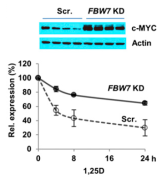 Ablation of FBW7 expression attenuates 1,25D-induced turnover of c-MYC expression |
| Vitamin D and the tumor suppressor FBW7. Notably, vitamin D signaling enhanced cMYC protein turnover and reduced that of MXD1, largely through controlling the function of the E3 ubiquitin ligase and tumor suppressor FBW7. FBW7 is a fascinating protein because it controls the proteasomal turnover of a wide range of proteins that drive cell proliferation. Our current efforts are focused on determining how the VDR controls FBW7 function and influences the turnover of multiple known and novel targets of FBW7. | |
Project 2. Chemical biology of vitamin D analogs (with Dr. Jim Gleason, Dept. of Chemistry).
We found a very strong synergistic effect between 1,25D and the histone deacetylase inhibitor (HDACi) TSA on 1,25D-resistant squamous carcinoma cells. While HDACs were initially characterized for their capacity to deacetylate histones, they are probably better known as protein deacetylases as some HDACs, notably HDAC6, are cytoplasmic and can deacetylate cytoplasmic proteins such as HSP90 and tubulin. HDACi's have been investigated for application in treatment of cancer. The anti-proliferative and cytotoxic activities of HDACi's have been demonstrated notably in breast, endometrial and ovarian cancer cells.
We collaborated with Dr. Jim Gleason in the Chemistry Department to develop and characterize a series of analogues of 1,25D, called triciferols (PNAS, 2008 [view pdf format]) that combine VDR agonism and HDAC inhibition. The parent compound displayed more efficacious antiproliferative activity in a series of cancer cell lines. In addition, it was more efficacious than 1,25D at inducing autophagic cell death in the MCF-7 breast cancer cell line. These compounds are rare in the annals of medicinal chemistry in that they are fully merged hybrid molecules that recognize two highly distinct targets; HDACs, and the VDR ligand binding domain. We have recently developed a series of more synthetically accessible non-steroidal hybrid compounds that display enhanced efficacy (Chemistry and Biology, 2012 [view PDF format]). We have now optimized both the VDR agonist and HDACi activities of these compounds and are testing the most efficacious in cancer cell line screens and animal models of cancer.
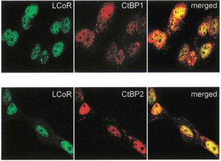
Project 3. Structure/function studies of LCoR, a corepressor of gene expression.We identified a novel nuclear factor that we called ligand-dependent corepressor (Mol. Cell, 2003 [view PDF format]). LCoR was identified as a protein that interacted with the ligand-bound estrogen receptor, which is a nuclear receptor and hormone-regulated transcription factor. This finding was remarkable as agonist-bound nuclear receptors usually recruit coactivators of transcription. LCoR represses ligand-dependent transcription by several steroid and non-steroid nuclear receptors. It binds to ligand-bound steroid receptors in vitro via a single “nuclear receptor” (NR) box, an amphipathic alpha helix containing the motif LXXLL. Mutation of this motif abolishes binding of LCoR to ligand-bound receptors, and disrupts its repressor activity. Mutagenesis of the estrogen receptor ligand binding domain identified a broad surface of interaction with LCoR centered on the coactivator binding pocket. |
|
|
LCoR is widely expressed in the adult and throughout fetal development. Highest levels are found in the placenta, where most of LCoR is expressed in the syncytiotrophoblast layer, a critical interphase of maternal and fetal circulation and an important site of steroid hormone signaling. Much of our effort on LCoR has focused on identifying its interacting partners. We found that LCoR functioned as a corepressor at least in part by recruiting HDACs and the corepressors CtBP (C-terminal binding protein) and its interacting partner CtIP (JBC 2009a and 2009b). More recently, we discovered that LCoR also interacts with other classes of transcription factors, notably the transcriptional repressor Kruppel-like factor 6 (KLF6) (JBC, 2012 [view pdf format]). Moreover, it also interacts with another corepressor Kruppel-associated protein 1 (KAP-1) and KAP-1-associated transcription factors to repress gene transcription (Nucleic Acids Research 2014). Our currents efforts are aimed at using high-throughput proteomics, bioinformatics and functional genomics to characterize the LCoR “interactome”. |
|