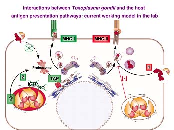
(1) Antigen presentation in the context of MHC-I in cells actively infected by Toxoplasma gondii
Primary astrocytes infected with transgenic T. gondii parasites secreting the model antigen ovalbumin in the vacuole. Blue (DAPI): astrocyte nuclei and parasite nuclear and apicoplast DNA. Green: complexes [MHC-I/OVA epitope] labeled by the specific monoclonal antibody 25-D1.16 (kindly provided by R. Germain).
Note that only infected cells present OVA in the context of MHC-I (Dzierszinski et al., in preparation).
(2) Manipulation of the MHC-II antigen presentation pathway and functional inactivation in cells infected by Toxoplasma gondii

Bone marrow-derived immature dendritic cells were first infected with transgenic T. gondii parasites expressing a cytosolic form of YFP (green: two parasites in a vacuole), and then received LPS as a signal of maturation. Red: surface and intracellular MHC-II molecules detected by immuno-fluorescence. Note that only uninfected cells show surface MHC-II staining and were able to mature as a response to LPS. We also showed that T. gondii infected immature DCs are unable to generate T cell activation.

Presentation of T. gondii antigen in the context of MHC-II actually requires parasite killing and phagocytosis. Splenic plasmacytoid DCs were purified from mice acutely infected with transgenic T. gondii parasites secreting the model antigen E alpha fused with RFP (red). Green: complexes [MHC-II / E alpha] labeled by the specific monoclonal antibody YAe (kindly provided by M. Jenkins). Note the morphology of the structure labeled in red, distinct from an actively formed vacuole.
(3) Control of Toxoplasma gondii encystation - Antigen presentation in immune privileged areas such as the CNS
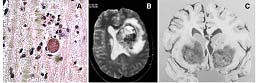
(A) T. gondii cyst in the brain of a chronically infected mouse (6 weeks post-infection). T cell infiltration is observed, but encephalitis is mild and parasite replication is controlled. (Image: F. Dzierszinski, unpublished.)
In immune deficient patients, parasite replication is not controlled and encephalitis develops. (B) Tomographic brain scan. (C) Bilateral areas of necrosis due to toxoplasmosis in basal ganglia and thalamus of a 67-year-old man.
Studying the biology of intracellular parasites is also a way of addressing various eukaryotic biological processes. Since the direct interactions occurring between antigen presenting cells, live intracellular pathogens and T cells shape the immune response against infectious organisms, these studies will provide new insights into T. gondii pathogenesis and will likely illuminate similar processes in other vacuolar pathogens. Investigating antigen delivery in the MHC presentation pathways in host cells infected with intracellular pathogens is expected to provide new elements for rational design of T cell vaccines and studies on immune surveillance of immuno-privileged sites.