

" Pathology is the basis of all true instruction in practical medicine.”
Samuel Wilks
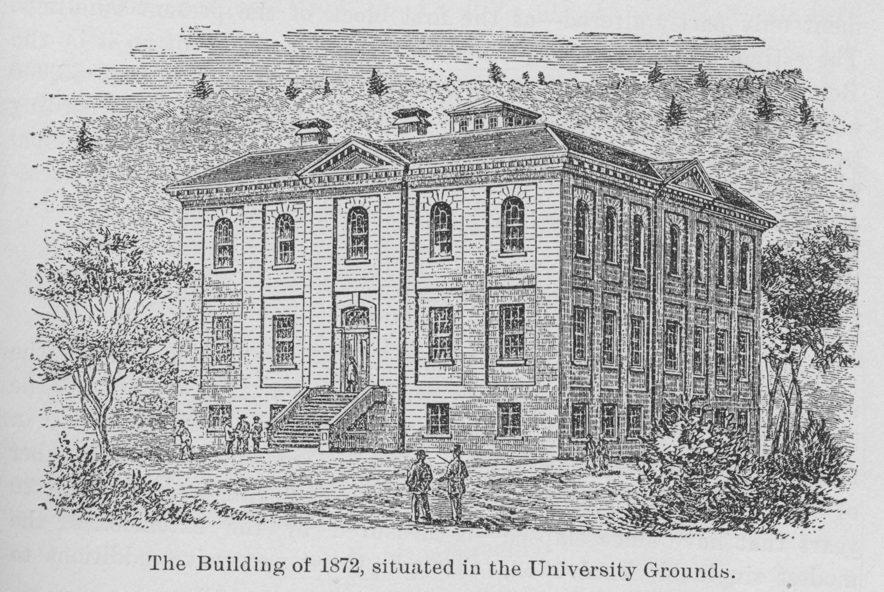
One of Osler's most important influences in the McGill Medical Faculty was on student teaching. In 1876, the McGill Medical Faculty announced a course called Practical Pathological Demonstrations to be given by Osler to the senior students. It was held in a lecture room in the McGill Medical Building for an hour on Saturday mornings and was based upon Virchow’s demonstrations which Osler had seen at the Berlin Pathological Institute in 1873.
Specimens which had been selected from autopsies performed at the Montreal General Hospital (MGH) the previous week were labeled, placed on trays and discussed by the students, with comments by Osler. The abnormalities seen in each specimen were correlated with the clinical history and notes that had been taken on the Hospital ward. Osler gave each student written descriptions of the specimens, usually four and sometimes as many as eight pages in length. Over 150 specimens were presented in this fashion during the 1877-1878 session.
In the 1878-1879 session, a second course entitled Instruction in Post-mortems was also listed in the University Calendar. Students performed autopsies in rotation under the supervision of Osler, again following the “method of Virchow” in which “system and thoroughness in inspection (were) insisted upon”. Osler clearly tried to impress upon his students the value of the autopsy in their medical education and it is likely that his enthusiasm for the process of clinico-pathologic correlation had a significant influence on their training.
"Dr Osler considered the post-mortem as the medical lesson par excellence, and he was always so anxious that the autopsies should be attended without fail, that he would not be satisfied with putting up a notice, but would tell his assistant, Dr. Wyatt Johnston, to advise us individually at the College, there was to be a post-mortem at such a time."
A. Schmidt (McGill 1886). Student Reminiscences, Montreal Period.
Bulletin IX International Association of Medical Museums and Technical Methods. 1926:183.
Osler’s pathology teaching also involved microscopy. Certainly, he had used the microscope while a student at McGill. Shortly after arriving in Montreal in 1874, Osler was hired to care for patients on the smallpox ward at the MGH. He spent the money he was paid for this work on 15 microscopes, which he used in a formal course on Practical Histology for the medical students in the summer of 1876. In this course, the students had to cut and stain their own tissue sections. Among the few of these that remain are examples of a number of pathologic abnormalities, including cancer, pneumoconiosis and tuberculosis. The details of how the students used these slides are unknown; however, it seems likely that they were derived from material Osler gave them from some of his autopsies.(See William Osler & the Teaching of Microscopy at McGill video at bottom of this page)
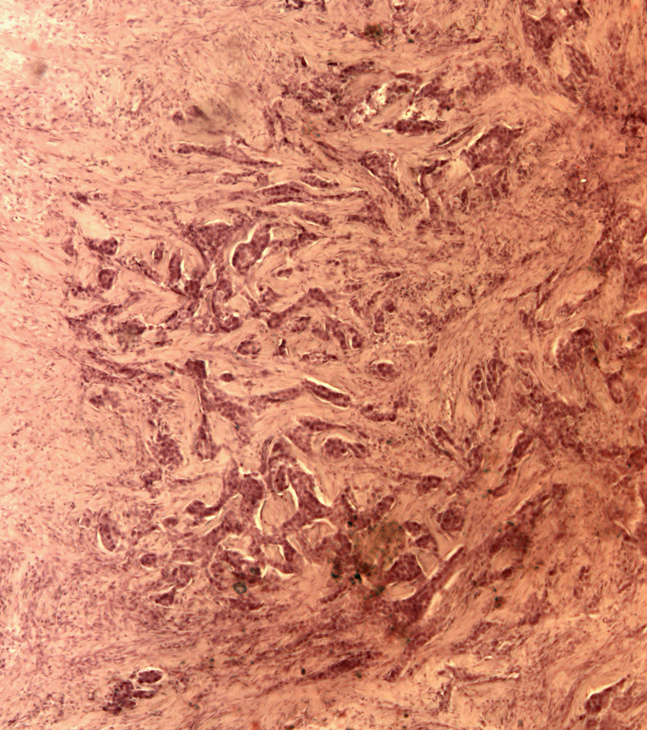
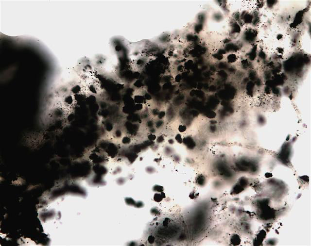
Image top: Invasive ductal carcinoma of the breast.
Image bottom: Anthracosis of the lung
These images were taken from slides prepared by Fred Green, a medical student at McGill from 1876 to 1878. They show an invasive ductal carcinoma of the breast (on the left) and anthracosis of the lung manifested by numerous macrophages stuffed with black carbon particles (on the right). The former is an example of the disease Osler discussed in his first pathological publication, Carcinoma mammae, (Canad Med J 1871-72;8:107-109) when he was a medical student in 1872. The latter is an example of “miner’s lung”, the subject of Osler’s first presentation at the Montreal Medico-Chirurgical Society in 1875 and of an extensive discussion in the Canada Medical and Surgical Journal of 1875-76 (Vol. 4 No. 4:145-169). Although neither slide can be definitely associated with the specific specimens Osler used for these demonstrations/publications, both were used at his 1877 summer session course in microscopy at McGill and show the same abnormalities. According to Abbott, gross specimens of the miner's lung were also present in the Museum in 1937*; however, they are now lost and presumably destroyed.
* Abbott, M. E. ‘‘More about Osler’’. Bull. Inst. Hist. Med., 1937, v, 787.
William Osler & the Teaching of Microscopy at McGill