QLS/CAMBAM Seminar - Dr. Paul Wiseman
Cellular cartography: Mapping protein transport and interactions in living cells with fluorescence imaging & fluctuation analysis
Paul Wiseman
McGill University
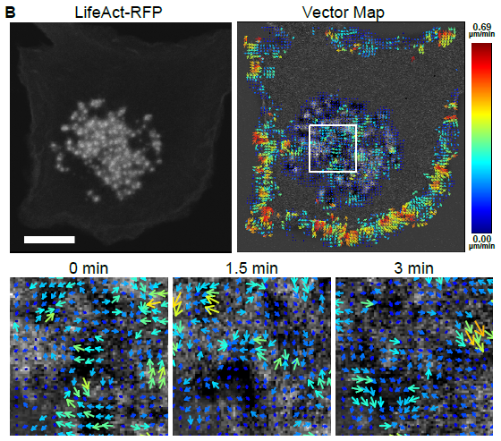
Image correlation methods are an extension of fluorescence fluctuation spectroscopy that can measure protein-protein interactions and macromolecular transport properties from input fluorescence microscopy images of living cells. These approaches are based on space and time correlation analysis of fluctuations in fluorescence intensity within images recorded as a time series using a fluorescence microscope. We previously introduced spatio-temporal image correlation spectroscopy (STICS) which measures vectors of protein flux in cells based on the calculation of a spatial correlation function as a function of time from an image time series.
Here we will describe the application of time window STICS and its two color extension, spatio-temporal image cross-correlation spectroscopy (STICCS), for measuring cellular waves of adhesion related macromolecules talin and vinculin as well as cytoskeletal actin between assembling and disassembling podosomes in dendritic immune cells. Podosomes are cylindrical membrane complexes with an integrin adhesive ring and an actin rich core that are associated with cellular migration and invasion in specific cell types. EM and super-resolution microscopy of cells shows radial actin filaments that connect neighboring podosomes so we applied pair vector correlation analysis to further characterize the transport waves within connected podosome clusters.
These analyses combined with pharmacological perturbation experiments show that podosome turnover is coordinated within local clusters in cells with a correlation length scale extending to next nearest neighbor podosomes. As well I will introduce future extensions of ICS including new extensions to super-resolution fluorescence microscopy.