
Our MEG program was inaugurated in 2012 and has become a busy unit of the McConnell Brain Imaging Centre. Our MEG users have published their data in top-tier journals and benefit from strong technical and software support from our core staff. We also have organised 4 week-long MEG training workshops, with attendees traveling from as far as Europe, China and Japan. Overall, our mission is to provide state-of-the-art support and expertise to everyone interested in learning or perfecting new skills in brain electrophysiology and imaging.
What is MEG?
Magnetoencephalography (MEG) is a neurophysiology modality for basic and clinical brain research at the scale of systems and networks. MEG measures non-invasively and in real time the tiny magnetic fields generated by neuronal currents. A unique asset of MEG imaging is its unrivaled temporal resolution, reaching the millisecond time scale across the entire brain. Clinically, MEG has been typically indicated for the pre-surgical work-up of severe, drug-resistant epilepsy and the functional pre-surgical mapping of brain tumors. There is also great potential in using MEG to investigate the pathophysiology of many other neurological syndromes and neuropsychiatric disorders (e.g., stroke, dementia, movement disorders, depression, Alzheimer’s disease, autism spectrum disorders, etc.). MEG has strong value in revealing the dynamics of brain activity in sensory perception, cognition and behavior: it has provided unique insight on the time-resolved processes of brain functions and network integration including resting-state dynamics. The MEG community comprises about 200 MEG centers worldwide. It is constantly contributing new methods and improving software tools to make the technique more accessible to a wider range of investigators.
Physiological origins of MEG signals

S. Baillet, Nature Neuroscience 20, 327–339 (2017)
Neurons generate electrophysiological currents (shown in yellow), which travel through the entire brain volume and head tissues. They eventually reach the scalp surface – although very distorted and attenuated – where they can be detected using electrodes with electroencephalography (EEG). Magnetic fields (in green) travel more freely across tissues and are much less distorted than current flows. They can be captured using arrays of magnetometers with MEG. The blue and red colors on the scalp illustrate the continuum of magnetic fields distributed at the surface of the head.
Scale of MEG signals
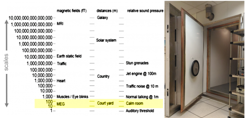
The ultra-sensitive MEG sensors deal with a range of environmental magnetic fields of about 10 to 12 orders of magnitude, most of which consist of nuisances and perturbations from external sources masking the brain activity at the femto-Tesla scale (1 fT is 10-15 T). A magnetically-shielded room (MSR) is therefore an important element to MEG sensing technology. It is built from a variety of metallic alloys meant to greatly attenuate external electromagnetic perturbations, including mu-metal, a nickel-iron alloy with a high magnetic permeability, which makes it very effective at screening external static or low-frequency magnetic fields. This makes MEG recordings possible, even in noisy environments like hospitals or nearby MRI scanners.
The MEG system

Courtesy CTF MISLP www.ctf.com
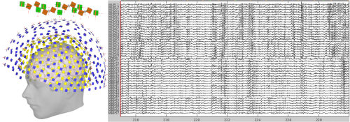
275 pick-up coils are arranged in a rigid helmet around the head. 64 channels of EEG can also be recorded simultaneously at up to 12 kHz sampling rate, i.e. one full brain image every 100 μs. The system can operate both in seated upright and supine horizontal positions, for maximum patient/participant comfort. The raw data consists of traces that represent the magnetic field fluctuations detected by each sensor.
Participant comfort and safety
The MEG instrument and environment are considered safe and there are no medical restrictions. The measurement is entirely passive: magnetic sensors inside a helmet measure the magnetic fields generated by neural activity that propagate outside the head. No energy, radiation or other agent is impressed, injected or used.
MEG requires minimal subject preparation: head-positioning coils are affixed to the skin to monitor head movements and standard ECG and EOG electrodes are positioned for basic monitoring of heart beats and eye movement respectively. The heart and eyes cause artefacts in MEG recordings and the electrode recordings help to attenuate them during analysis. All equipment and supplies for a generic MEG session have received adequate safety certifications for human use.
The MEG instrument is located inside a passive magnetically shielded room (MSR), consisting of 3 layers of metal alloys to attenuate magnetic interference from the environment. The room is about 80 sq ft. At all times, participants can communicate via video and audio intercom with the operators outside the room. A caregiver can stay in the MSR seating next to the participant, if necessary. They can also exit the room at all times by operating the hand wheel from inside the MSR.
Participants sit or lie down on a comfortable chair/bed. Unlike an MRI "tunnel", only their head is brought inside the rigid MEG helmet, which covers essentially the lateral, superior and posterior aspects of the cranium, leaving the face and eyes unobstructed. This is usually well tolerated by participants prone to claustrophobia. There is no noise generated during acquisition, hence MEG is generally considered comfortable to participants, including for most patients and young children.
Another potential risk from MEG is the spontaneous boil-off of the liquid helium used for refrigeration of the superconducting sensing system. This is similar to MRI quenching, though the volume is much smaller in an MEG system. Although very unlikely, boil-off can occur with minimal risk to participants: the MEG instrument has safety valves to prevent pressure from building inside the helium reservoir and if helium is released, it would rise to the ceiling and be vented rapidly by the air ventilation system inside the MSR. Although helium is inert, there is a risk of oxygen depletion inside the MSR in case of massive helium release. Because of this, there is an oxygen sensor inside the MSR and an alarm would sound if low levels are detected. It would take only seconds for the attending personnel to open the MSR door, which would immediately resolve the situation.
Finding task-related brain activity
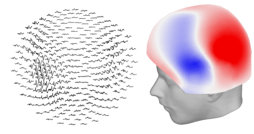
There is constant neuronal activity all across the brain. Therefore to separate signals related to a specific function from this background activity, we often need to average the signal over many repetitions of the same task. The images on the right show the average signal recorded when a subject hears a tone, over 200 trials. The left image shows the average traces at each sensor location, and the right image shows the average field distribution at 100 ms after the tone onset.
Registration with MRI

The 3-D locations of anatomical fiducial points are digitized to overlay the MEG results on high-resolution MRI images. Head-positioning indicators are also taped to the subject’s head to detect and compensate for small head movements occurring during the session.
Merging anatomy and function

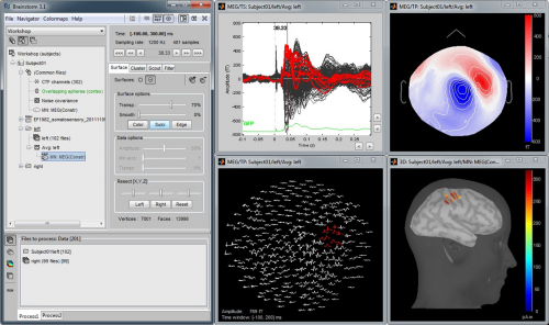
MEG functional data is combined with MRI structural information. Head tissues are identified and extracted from the MR image volume and are used to model the electromagnetic properties of the head (left) to localize the brain regions that have generated the MEG signals. The images on the right show the average brain response to a subject hearing a tone 200 times.

Research activities
Our core research activities focus on developing new methods, analysis techniques and software to advance MEG as a functional brain imaging modality.
Cross-frequency interaction: Brain rhythms are coupled and interdependent, which can be revealed by measures such as phase–amplitude coupling (PAC). Here, PAC analysis of MEG traces obtained in the resting-state of a healthy adult shows that the amplitude of fast gamma activity is modulated by the phase of slower oscillations.

E. Florin & S. Baillet, The brain’s resting-state activity is shaped by synchronized cross-frequency coupling of neural oscillations, NeuroImage 111, 26–35 (2015)
Neurofeedback: We have developed the technology to use MEG as a real-time, closed-loop imaging technique. Real-time reconstruction of brain activity is performed as the subject is being scanned. Indices of activity/connectivity of key brain areas are extracted and presented back to the subject. The goal is to promote predefined patterns of brain activity, which are designed for a determined objective. Some clinical applications are on the horizon: epilepsy seizure control, optimized motor and speech rehabilitation after stroke, etc.

G. Sudre et al., rtMEG: a Real-time Software Interface for Magnetoencephalography, Comput Intell Neurosci, 327953 (2011)
Connectivity: Brain regions communicate very rapidly with one another, forming networks that contribute to the integration of information by the brain. MEG can help reveal how these regions communicate. The figure to the right represents virtual axonal white fibers estimated from functional connectivity metrics obtained with MEG source imaging. Three different frequency bands are represented showing the brain regions connected with the strongest coupling in those bands.

Sébastien Déry
Clinical applications
Presurgical mapping of drug-resistant epilepsy: Regions involved in the early onset of epileptic events can be detected thanks to the millisecond time resolution of MEG. Additionally, the localization of crucial brain functions (language, motor control) can be mapped on the patient’s brain and help determine the best target for neurosurgery. The image on the left shows fast propagation of epileptic activity detected with MEG source imaging, shown in red, as registered on the patient's anatomy obtained with MRI structural imaging.

Presurgical mapping of brain tumors: MEG can help determine whether brain regions within or in the vicinity of a malignant brain tumor are involved in critical brain functions such as language and motor processes. Combined with other imaging modalities, this information can assist neurosurgeons in planning the safest surgical approach on an individual basis. The figure shows time-resolved mapping of language-related processes (in orange) in a brain tumor patient during a word-reading test. The lesion is shown in green.

Collaboration with Binder's Language Laboratory (Medical College of Wisconsin).
Please see BIC Booking & Pricing for policies and fees for all BIC facilities.
This section provides additional details specific to the MEG Unit.
New Studies
For scientific review for a new MEG study, please send the full details of the protocol, as you would for ethics review, to the sylvain.baillet [at] mcgill.ca (subject: New%20MEG%20study%20scientific%20approval) (MEG Unit Director). There isn't a specific required form to use for this, e.g. it can be based on an ethics application form or scientific review form for another committee.
You may use these examples and templates to help prepare your scientific and ethics review submissions.
- MEG Study Protocol Example
- MEG Ad English / French
- MEG Declaration of Consent English / French
You are invited to contribute your data to the Open MEG Archive (OMEGA). The goal of the OMEGA is to build and share a large collection of anonymized MEG recordings of spontaneous brain activity and brain responses to basic stimulation. This repository is made available to the academic community and may help researchers gain a better understanding of the mechanisms underlying brain activity, which may impact the discovery of new markers of neurological diseases or psychiatric conditions.
Instrument and Staff Availability
The MEG instrument is available 7 days a week / 24 hours a day to investigators with MEG-operator certification (see Training).
The MEG staff is available to assist investigators for MEG measurements or obtaining certification. Regular hours are Monday to Friday, 9:00 -17:00.
System maintenance imposes interruptions of MEG measurements for one hour once or twice a week, usually at the end of the day, Wednesdays and Fridays. The MEG staff will accommodate booking on these days to the best of their ability.
The time necessary for subject preparation varies from a minimum of 15 minutes for a MEG-only acquisition to an hour when EEG needs to be acquired simultaneously with MEG. The recording time depends on the protocol and will vary by study. If additional time is used, it will be billed (see BIC Facilities Billing).
MEG Data Collection and Retrieval
Raw data is collected and stored on the acquisition workstation (quickly.bic.mni.mcgill.ca). Typical disk space usage are about 10 GB per hour of recording. The raw data is then transferred overnight to the BIC cloud data storage (box.bic) and made available to their respective investigator for at least a month following the session date. Data is automatically deleted each month. It is therefore the sole responsibility of the investigator to retrieve and verify their data in a timely manner.
For investigators who have access to the BIC data storage, raw MEG data can be copied directly to that storage space. Arrangements must be made directly with the MEG system manager. More information regarding access and billing rates for BIC data storage is provided under Neuroinformatics.
Billing
For McGill users: you may use the Activity code 010243 (MEG Scanning) in your FOAPAL.
Returning researchers are billed hourly, with a discounted rate for certified MEG operators.

MEG-Infinity
The MEG-infinity program is a subscription-based billing model for researchers new to the MEG Unit. The program enables high-powered neuroscience research, encourages the adoption of open-science practices, and maximizes the impact of research funds.
MEG-infinity offers unlimited 24/7 access to MEG scanning against the payment of a flat subscription fee, valid over a 6-month period from the first booking. The subscription is for one study, with an unlimited number of participants (cross-sectional design) and/or sessions (longitudinal design).
The rates apply to MEG certified users who have been trained and approved to operate the system without assistance (see Training tab). The certification program requires 3 hours of supervised data collection, from the study of interest, at the regular hourly rate.
Studies that require full assistance from our MEG staff for data collection remain at the hourly rate. Current BIC cancellation policies apply, with special attention against booking-cancelling several blocks of time at once.
275-channel CTF MEG 2005 Series
- Manufacturer: CTF MEG International Services Limited Partnership (CTF MISLP)
- 275 axial gradiometer SQUID channels in a head-shaped helmet, up to 12 kHz sampling rate
- 29 reference channels for reducing environmental magnetic signals
- 64 simultaneous EEG channels (56 + 8 bipolar)
- 16 analog input channels,
- Digital input channels: DB37, two parallel (DB25) and two serial (DB9) ports
- Continuous head position tracking
- Located in a magnetically shielded room
- Comfortable reclining chair or bed for seated or supine participant positioning
Auxiliary devices
- VPixx ProPixx projector (1920 x 1080 resolution, up to 500 Hz color, 1440 Hz greyscale)
- E-A-RTone insert earphones with disposable foam tips
- Two Digitimer DS7A constant current stimulators
- Eye tracking (VPixx)
- Various response devices: VPixx ResponsePixx MRI system with two custom 5-button pads, finger tapper, MEG-compatible USB keyboard and mouse, joystick
- Audio & video participant/patient monitoring and recording capabilities
Stimulus software
- Matlab & PsychToolbox
- Presentation
- E-Prime
- OpenSesame
- PsychoPy
- Real-time neuro-feedback with Brainstorm
Investigators that collect MEG data can use the workstations in the MEG suite to learn how to pre-process and analyze their data with the assistance of the MEG team. This access is typically granted for a period of time, pending availability of workstations, long enough to analyze 1-2 subjects and develop an analysis workflow.
For investigators looking for MEG training or that are interested in learning more about Brainstorm, there are several training courses offered throughout the year.
Obtaining MEG Operator Certification
Investigators are encouraged to obtain certification to become an operator of the MEG instrument to benefit from 24/7 access to the MEG instrument, i.e. access outside regular opening hours and a discount on rates.
To obtain certification, the investigator should contact the marc.lalancette2 [at] mcgill.ca (MEG System Manager). Training begins with one or two sessions (about 3 hours) in the MEG lab to cover many aspects:
- subject safety,
- equipment operation,
- basic system design and principles,
- general procedure for participant set up and data collection,
- acquisition software,
- identifying artefacts,
- troubleshooting.
Following this, the trainee will be supervised and assisted by the MEG System Manager during at least the first 3 recording sessions of his/her own study. These training sessions are billed at the regular data acquisition rate (with assistance). There is no additional cost to obtaining this certification.
The MEG System Manager and the Director of MEG Research may require that additional training sessions are necessary before obtaining certification. Additional training sessions can also be requested by the investigator.
The investigator will then be notified by a letter from the Director of MEG Research that he/she has obtained certification to operate the MEG system independently. The certification is valid for all current and future studies run by this investigator. The MEG Program will maintain a list of certified MEG operators so that other PI's are encouraged to use their services.
Our Team
We offer expertise, training and support to all investigators interested in learning about MEG and performing a MEG study. We also develop our own research in the neuroSPEED laboratory.
| Dr Sylvain Baillet | Director, MEG Unit |
| marc.lalancette2 [at] mcgill.ca (Marc Lalancette) | MEG System Manager |
| raymundo.cassani [at] mcgill.ca (Raymundo Cassani) | Core Software Developer |
For inquiries and more information, please contact the Director of the MEG Unit.
Software
The analysis and interpretation of MEG data require the exploration of large volumes of data. Our program contributes original software developments to facilitate the integration of MEG in the standard clinical workflow and promote research productivity.
Our main effort is Brainstorm, a collaborative, open-source application dedicated to the analysis of multimodal electrophysiology, with the objective of sharing a comprehensive set of user-friendly tools with the scientific community. For physicians and researchers, the main advantage of Brainstorm is its rich and intuitive graphic interface, which does not require any programming knowledge, a rich online documentation and strong user support (20,000 user accounts, 950 journal articles, 36 training workshops since 2011).
Brainstorm Consortium: McGill, USC, Cleveland Clinic, CNRS/INSERM (France); NIH funded.
Open Science
Our unit is also at the forefront of open-science efforts. The Open MEG Archive (OMEGA) is the fruit of a collaborative effort between our program and the Université de Montréal to provide a core repository of MEG data for open dissemination. It now contains multimodal data from over 200 participants, about 300 resting-state MEG records. It also features anatomical T1-weighted MRI volumes and anonymized demographic and questionnaire data. OMEGA will continue to expand, with contributions from the scientific community.


The Brain Imaging Data Structure (BIDS) is an emerging standard for the harmonization of neuroimaging data, to which we contributed to extend to MEG. The MEG-BIDS specifications were defined in accord with best-practice guidelines for conducting MEG research, with direct inputs from MEG investigators, technical support staff and data managers. All MEG data collected at our unit is automatically organized according to BIDS when copied from the acquisition system to our storage server, where researchers access their recordings.